What Cord Blood Research Shows About Muscle Repair
Mesenchymal stem cells from cord blood activate muscle's own repair machinery.
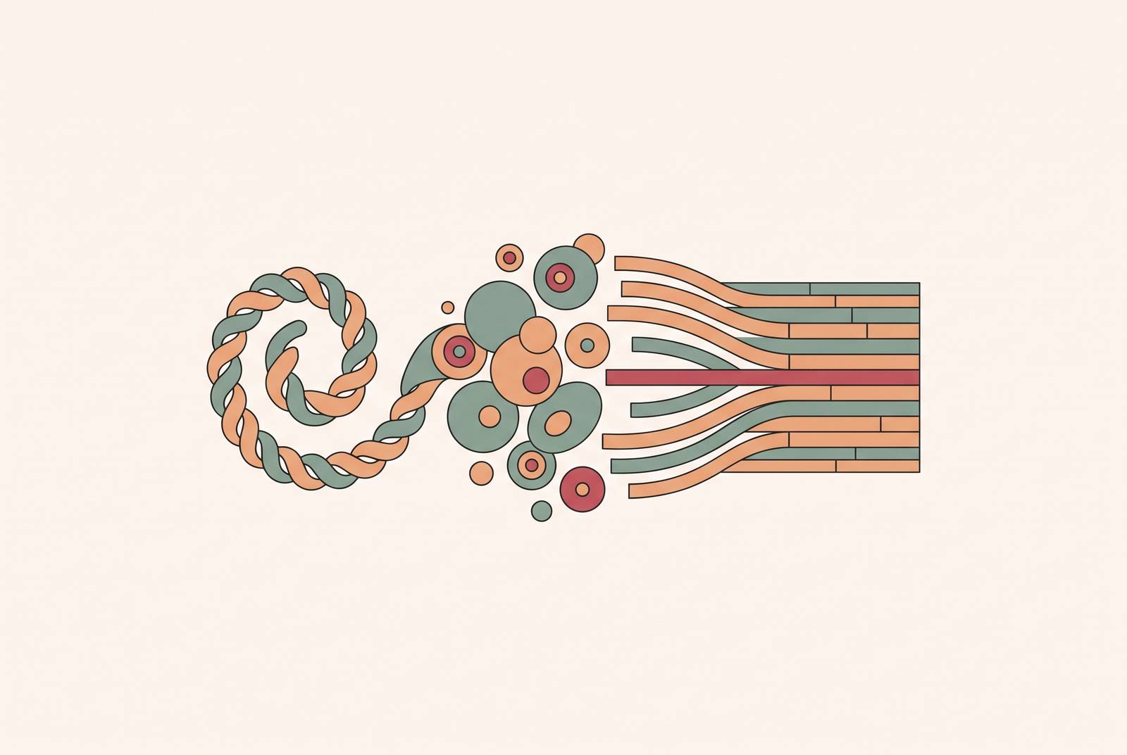
Cord blood is not one thing. It carries multiple distinct cell populations, and only recently has research started sorting out which of them matter for muscle repair, and why. This piece traces what that research actually shows, from lab dish to animal model to a handful of early human trials, and it's blunt about where the claims outrun the data. Mesenchymal stem cells (MSCs) do the real work here, and anyone treating cord blood as one interchangeable product, easy to swap for another, is missing what actually sits in the vial.
Hematopoietic stem cells (HSCs) are the population most people think of when they hear "cord blood banking," and for good reason: they rebuild blood and immune systems, and they've been the clinical workhorse for decades with real transplant history behind them. But HSCs aren't the muscle story. That role belongs to MSCs, a multipotent population that turns into bone, cartilage, and muscle tissue, and one that also carries strong immune-signaling functions. A 2025 review in PMC called umbilical cord MSCs a "clinically attractive regenerative and immunomodulatory platform that combines ethical accessibility, low immunogenicity, rapid expansion, genetic stability, and a potent paracrine secretome." Strip the jargon and it names four plain advantages: the cells come out without harming the donor, they exist in workable quantities, they freeze well for long-term storage, and because they come from newborns, they trigger less immune rejection than adult cells when given to someone else. HSCs and exosome-secreting cell populations play supporting roles too, but the muscle repair story runs through MSCs first.
How skeletal muscle normally repairs itself and where that process breaks down
Muscle regeneration runs in three phases: destruction, repair, and remodeling. Each phase has its own cast of cells, and where cord blood-derived therapy might step in depends on how those cells hand off to each other.
Destruction comes first. An injury creates a dead zone marked by a contraction band, condensed cytoskeletal material left behind where the cell gave out. The sarcolemma, the muscle fiber's outer membrane, ruptures and spills its contents into the bloodstream. That damage hits nearby blood vessels too, which sets off both the coagulation cascade and the complement system. Local immune cells sense the damage and fire off inflammation almost immediately. None of this is optional. It's simply how muscle signals that it's hurt.
Repair depends on satellite cells, muscle's built-in stem cell reserve. These cells sit quietly between the plasma membrane and basal membrane of each fiber until an injury wakes them. Once activated, a satellite cell divides asymmetrically, producing myoblasts that multiply, differentiate, and fuse into new multinucleate, contractile fibers. The whole sequence runs under a strict order of proteins switching on one after another: Pax7, then Myf5, then MyoD, then Myogenin, then MRF4. If any link in that chain stalls, the fiber never finishes forming.
Not every injury heals the same way, and this is where a lot of the optimism about cell therapy needs a reality check. Some forms of acute injury, where the basic scaffold of the muscle remains intact, can heal with meaningful fidelity. Volumetric muscle loss doesn't work that way: when a chunk of muscle is physically gone, nothing grows back on its own, and no clinical treatment so far restores full function after that kind of loss. Chronic diseases like Duchenne muscular dystrophy, sarcopenia (age-related muscle wasting), and myasthenia gravis fail for a related but distinct reason. Either the satellite cell pool itself runs out after repeated cycles of damage, or the surrounding inflammation drowns out whatever regenerative signal is trying to get through.
That failure point, satellite cells stalling or inflammation swamping the repair signal, is exactly where cord blood-derived MSCs have a mechanistic argument for stepping in.
How cord blood MSCs differentiate directly into muscle cells
Under the right lab conditions, cord blood MSCs walk through the same sequence of transcription factors that satellite cells use on their own. Researchers inducing myogenic differentiation have tracked the timeline directly: Pax7 and MyoD show up around day 3, marking early muscle progenitor identity. Myogenin appears by day 6, the commitment point toward becoming muscle rather than something else. By day 10, myosin heavy chain, the actual contractile protein that makes muscle contract, appears in the cells. Flow cytometry and immunofluorescence imaging confirm each stage.
Disease-specific work backs this up with a sharper result. CD34+ cord blood cells placed in co-culture with Duchenne muscular dystrophy myoblasts start fusing with them almost immediately, and by day 15 the fused cells mature into full myotubes. Some of those CD34+ cells went further and started making dystrophin, the exact protein DMD patients are missing. That held up both in the dish and in earlier animal work, and it matters because dystrophin production is the actual therapeutic target in DMD, a direct measure of the therapy's effect rather than a proxy for general muscle health.
Cardiac muscle tells a related story. A University of Bristol study, funded by the British Heart Foundation and the NIHR and published in Stem Cell Reviews and Reports in 2011, found that CD133+ cord blood stem cells could be expanded up to sevenfold in the lab and then coaxed into cardiac muscle-like cells. That's a different differentiation path than skeletal myogenesis, but it points to the same underlying fact: cord blood progenitor cells aren't locked into one tissue fate.
The complication deserves sitting with rather than glossing over. A study in Scientific Reports found that umbilical cord tissue, specifically Wharton's jelly, outperforms cord blood as a source of MSCs for turning into muscle. On that one measure, direct differentiation, cord tissue wins outright, and pretending otherwise does the research no favors. Cord blood's strongest card lies elsewhere, in becoming muscle only weakly by comparison. Its value sits elsewhere, in immune signaling and paracrine communication, which the next two sections cover.
MSC activation of the muscle's own satellite cells rather than replacement of them
Treating differentiation as the main job of MSCs undersells what they actually do, and the evidence points the other way: their bigger contribution is waking up cells that are already there. According to a review in Frontiers in Cell and Developmental Biology, MSCs rouse the muscle's existing satellite cell population, supporting the tissue's own regenerative response rather than replacing it. That's a different mode of action than direct replacement, and the more useful one, since it puts the muscle's own repair machinery back to work instead of trying to substitute for it.
Research on myasthenia gravis, an autoimmune disease that attacks the neuromuscular junction, shows this mechanism working in practice. Human umbilical cord MSCs remodeled the extracellular matrix around damaged muscle, raising levels of laminin and collagen IV, structural proteins the tissue needs to rebuild scaffolding. At the same time, they activated the satellite cell program directly and improved signaling at the neuromuscular junction itself. MSC activity also ties into acetylcholine metabolism, the chemical messenger nerves use to talk to muscle, which matters for MG specifically and for any condition where that nerve-muscle interface breaks down.
Age adds another layer. As people get older, their own MSC population shrinks, and that decline speeds up fat infiltration into muscle while weakening regenerative capacity across the board. That's a real argument for why neonatal, cord blood-derived MSCs might have a wider therapeutic window than a person's own adult stem cells, which have already aged along with the rest of the body. But activation only works if a satellite cell reserve is still there to wake up. In advanced disease, once that reserve is gone, this approach hits a real ceiling, because it functions only as a support mechanism.
The anti-inflammatory role that protects muscle from its own repair response
Inflammation is necessary early in muscle repair, but only briefly. Neutrophils rush in right after injury, and that's normal, part of clearing debris. But if that neutrophil response doesn't shut off on schedule, the tissue drifts toward fibrosis, scarring, instead of regeneration.
A mouse study using bupivacaine injections into the quadriceps of C57BL/6 mice showed how engrafted Wharton's jelly-derived MSCs interrupt that drift. Treated mice showed a near-total shutdown of neutrophil infiltration, driven by a drop in the chemical signals that normally recruit neutrophils to the site: C5a, KC, MIP-2, LIX, Fractalkine, LTB4, and interferon-gamma. Less fibrosis followed, and so did better functional recovery.
Autoimmune disease shows a parallel effect. In experimental autoimmune myasthenia gravis models, MSC transplantation lowered the abnormally high levels of acetylcholine receptor antibodies driving the disease, and it restored the balance between Th17 and regulatory T cells, two immune cell types normally at odds with each other. Research published in Frontiers in Cell and Developmental Biology ran MSCs alongside human peripheral blood mononuclear cells in a humanized mouse model of MG and cut disease severity in half within two weeks of injection. Two weeks, for a disease that typically only worsens without intervention, is fast.
This matters past autoimmune disease specifically. Duchenne muscular dystrophy and sarcopenia both carry a chronic, low-grade inflammatory backdrop as part of the disease process itself, a core driver of the disease rather than a side effect of it. MSC-driven immune regulation goes after that underlying driver directly, instead of only patching the damage it causes. Cord blood MSCs bring a practical edge here too: their low immunogenicity means they can be used allogeneically, in someone other than the donor, without the heavy immune suppression that limits so many other cell-based treatments.
The contribution of exosomes and paracrine signaling to the repair picture
Much of what MSCs accomplish, they accomplish without ever becoming muscle cells. Instead, they secrete a stream of pro-regenerative signaling molecules, and exosomes are the main delivery vehicle for that signal.
Cord blood-derived exosomes are small extracellular vesicles marked by surface proteins CD9, CD63, and CD81. Inside, they carry proteins, lipids, microRNAs, and signaling lipids, and recipient cells take up that cargo either through direct membrane fusion or through phagocytosis and endocytosis. That cargo carries signaling molecules that influence the cellular processes driving fibrosis, positioning exosomes as potential modulators of scar tissue formation.
A sarcopenia study using SAMP10 mice, a strain of 24-week-old male mice bred to model accelerated human muscle aging, gave the animals exosomes from human umbilical cord tissue intravenously. Twelve weeks later, treated mice showed better skeletal muscle structure and performance, along with higher levels of proteins tied to muscle maintenance and energy metabolism: p-mTOR, myosin heavy chain, PGC-1α, and Sirt1.
Exosomes carry practical advantages living cells don't. They provoke less of an immune response, and production and storage can be standardized in ways whole-cell transplants can't easily match. More than 40,000 PubMed publications now cover exosomes, and over 100 clinical trials are testing them across different diseases, so the interest here isn't fringe. For cord blood specifically, cell-free exosome therapy offers a way to deliver the biological signal without needing the original cells to survive and engraft, which matters for anyone thinking about cord blood banking as a long-term resource.
What the research shows across specific muscle conditions
Duchenne muscular dystrophy is an X-linked disease that damages proximal skeletal muscle, respiratory muscle, and heart muscle over time. Human umbilical cord MSCs have shown measurable benefit in DMD research: better motor function, more muscle strength, and lower blood levels of creatine kinase, lactate dehydrogenase, ALT, and AST, all markers that climb when muscle is actively breaking down. A registered trial, listed as NCT01610440, was built specifically to test the safety and effectiveness of hUC-MSC transplantation in DMD patients. And the earlier fusion experiment, where CD34+ cord blood cells fused with DMD myoblasts and produced dystrophin, still stands as proof that correcting the underlying genetic defect isn't out of reach.
Myasthenia gravis is fundamentally an immune disease that happens to show up as muscle weakness, since the immune attack targets the neuromuscular junction directly. MSC therapy in MG research addresses both ends of that problem at once: it reduces the antibody levels driving the attack, rebalances the Th17/Treg ratio, and supports the muscle tissue weakened as a downstream consequence. The 2025 humanized mouse study published in Frontiers in Cell and Developmental Biology, again, cut disease severity in half within two weeks.
Sarcopenia is driven by a cluster of overlapping problems: chronic low-grade inflammation, more cell death, mitochondrial dysfunction, and a regenerative process that slows with age. Research into umbilical cord MSCs has found that they suppress cell death, calm inflammation, support new blood vessel growth, and remodel the extracellular matrix in aging muscle. The SAMP10 exosome study backs that up with a concrete functional readout at 12 weeks.
Volumetric muscle loss remains the hardest case, and no one studying it claims otherwise. When muscle mass is physically gone, nothing grows back on its own, so combination approaches are the current frontier. One study paired human umbilical cord MSCs with a decellularized porcine cardiac extracellular matrix scaffold in a rat hindlimb model. By 8 weeks post-injury, the combined treatment group showed better regeneration, better functional recovery, and less collagen buildup than other arms in the study, published in the Journal of Orthopaedic Research in 2019. Even here, the limiting factor is that MSCs don't stick around long in injured tissue once transplanted. The scaffold compensates for that some, but it's a partial fix, not a full one.
Cardiac muscle research runs on a related but separate track. The University of Bristol's 2011 work with CD133+ cord blood cells, expanded sevenfold and then differentiated into cardiac muscle-like cells, points to a specific advantage of the cord blood source. Adult stem cells harvested after a heart attack tend to be less functional simply because they've aged along with the patient, while cord blood cells sidestep that problem by coming from birth.
The state of the clinical evidence and its unproven limits
Most of what's described above comes from animal models, dish-based differentiation experiments, and co-culture studies. The underlying biology is mapped out in real detail at this point. The human clinical evidence sits much earlier, and that gap is the honest headline here, not a footnote to it. State what the data will bear: the mechanism is proven, the treatment isn't, and conflating the two is the single most common mistake in how this research gets talked about.
Where human trials exist, the functional gains reported are real but modest, and they vary a lot by condition. Sarcopenia research has reported measurable improvements on functional outcome measures, a meaningful signal but not a dramatic one. DMD research, including the HOPE-2 trial, has shown a slowing in the rate of upper-limb functional decline rather than a reversal of it. Volumetric muscle loss work using scaffolds seeded with cell therapy has produced better regeneration in animal models, but nothing close to full functional restoration, and that gap between animal result and human outcome hasn't closed.
None of that undercuts the mechanistic case built across differentiation, satellite cell activation, immune modulation, and exosome signaling. Those pathways are documented, specific, and reproducible across multiple labs and models. But mechanism and clinical proof are different things. Cord blood-derived cells have several distinct, well-characterized ways of supporting muscle repair, and that much is settled. Whether that mechanistic promise becomes an approved, reliable treatment is a question the current trials haven't answered, and no amount of promising cell biology in a dish changes that fact.
Sources
- Stem cells from cord blood could help repair damaged heart muscle
- Frontiers | Restoration of skeletal muscle function via mesenchymal stem cells: mechanistic insights and therapeutic advances in myasthenia gravis
- Advancing regenerative therapies with umbilical cord-derived mesenchymal stem cells: A review - PMC
- Adult stem cells at work: regenerating skeletal muscle - PMC
- nature.com
- pmc.ncbi.nlm.nih.gov