Neonatal Iron Transfer Physiology During Cord Clamping
Delayed cord clamping preserves fetal iron transfer through a narrowing physiological window.

The timing of cord clamping governs how much iron a newborn carries into the first year of life, and that claim only makes sense once the delivery mechanism behind it is clear. The placenta is not a passive membrane that lets iron seep through by concentration gradient. It runs an active transport chain, and every link in that chain determines how much iron reaches the fetus before the cord is ever touched.
Iron moves through maternal blood bound to transferrin, and the placenta grabs it with transferrin receptors that sit on the brush border membrane, where maternal circulation touches it directly. Once bound, the transferrin-receptor complex gets pulled into the placental cell inside coated vesicles. Those vesicles are then acidified, a chemical step that strips iron free and releases it into the cytosol. Divalent metal transporter-1, known as DMT1, partially governs that release, acting as one of the gatekeepers that decides how efficiently iron moves out of the vesicle and into usable cellular space. From there, iron has to cross again, this time out of the placental cell and into fetal circulation, a handoff carried out by ferroportin-1 working alongside a placental copper oxidase.
Each step in that chain, transferrin receptor capture, vesicle acidification, DMT1-mediated release, ferroportin export, is a rate-limiting point. Slowing down or disrupting any single one drops fetal iron uptake, regardless of how much iron is circulating in the mother's blood. Pregnancy builds the fetal iron reserve gradually, across months. By the time labor begins, the fetus has been accumulating iron through this exact pathway for the better part of gestation. The cord at delivery is the final leg of a transport relay that has been running since early pregnancy, and what happens to that cord in the minutes after birth either completes the relay or cuts it off mid-handoff.
Umbilical vessel behavior at birth
The cord contains three vessels, two arteries and one vein, and the timing difference in how they respond to birth is the physiological basis for why the moment of clamping matters.
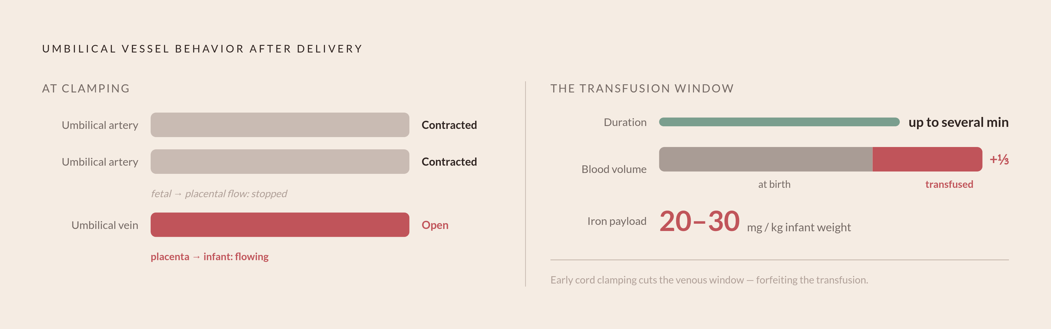
The two umbilical arteries, which carry blood from the fetus back toward the placenta, contract first. Within moments of delivery, arterial flow drops sharply, cutting off the fetal-to-placental leg of the circuit. The umbilical vein behaves differently. It carries oxygenated, iron-rich blood from the placenta toward the newborn, and it can keep that flow moving for up to several minutes after birth, even as the arteries have already begun shutting down. The circuit becomes one-directional: blood still moves from placenta to infant through the vein, with little or nothing moving back the other way.
That one-way venous window is precisely what delayed cord clamping is designed to preserve. For as long as the vein stays open, more blood keeps moving from placenta to infant, up to roughly a third of the newborn's final blood volume. That blood is not generic volume. It carries red blood cells, plasma proteins, stem cells, and an iron payload, and that payload comes to about 20 to 30 mg for every kilogram the infant weighs. That iron does not sit inert in the bloodstream. It gets folded into neonatal circulation fast, and then it turns into stored ferritin, the form the infant's body draws on for months after birth.
Clamping the cord early does not happen because placental iron transport has finished. It happens because the clinician has physically interrupted a process that was still active. The arteries closing on their own is biology. The vein closing because of a clamp applied at ten seconds instead of three minutes is a decision, and it is a decision with a measurable iron cost attached to it.
The placenta's response to low maternal iron
When a mother's iron stores run low, the placenta does not just pass along a smaller share of what little iron is there. Instead, it upregulates its own transport machinery, so it works harder to pull every available unit of iron across to the fetus. That compensation has a ceiling, and where that ceiling sits determines how much weight falls on the cord-clamping window at delivery.
You can see this upregulation at both the mRNA and protein level, across the whole transport chain described earlier. Transferrin receptor expression rises in iron-deficient placentas, a pattern also observed in BeWo cell models used to study placental transport in the lab. DMT1 levels rise alongside it, increasing the rate at which iron moves into the syncytiotrophoblast, the placental cell layer in direct contact with maternal blood. The placenta, in effect, is extracting more iron per unit of maternal supply than it would under normal conditions.
That compensation helps, but it does not close the gap. Fetuses born to iron-deficient mothers still carry elevated risk of developmental delay, neonatal mortality, and broader morbidity. Placental upregulation can partially offset a shortfall in maternal supply, but it cannot replace the supply that was never there to begin with. For these mother-infant pairs, the venous window at delivery no longer adds to an antenatal transfer that was already enough. It becomes a disproportionately large share of whatever iron endowment the infant is going to get. Early clamping in this context is not a neutral choice applied against a full reserve. It compounds a deficit that began months earlier in utero.
The scale of this problem is not marginal. A 2026 global review finds that iron deficiency anemia affects roughly half of pregnant women in developing countries, and it is highest in Asia. The population for whom the cord's venous window matters most overlaps heavily with the population that has the least room to correct an iron shortfall through diet after birth.
What early cord clamping cuts short in hematological terms
The consequences of early clamping are not theoretical. They show up as measurable differences in hemoglobin, hematocrit, and stored iron, differences detectable within days of birth and significant enough to track in clinical research.
A 2025 prospective observational study found significant differences between early cord clamping and delayed cord clamping groups across mean hemoglobin levels, hematocrit values, birth weight, and five-minute Apgar scores. Delayed clamping increased neonatal hemoglobin concentration at birth and improved iron reserve during early infancy. A separate prospective observational cohort tracked hemoglobin and bilirubin across both preterm and term infants under different clamping timings, producing paired data that lets researchers compare what early versus later clamping yields in the first days of life across multiple populations.
What gets lost in early clamping does not get made up quickly afterward. Iron stores built through placental transfusion are what carry an infant through the first several months of life, when breast milk alone gives little iron and solid, iron-containing foods are not yet part of the diet. An infant who misses out on the placental transfusion at birth is drawing down a smaller reserve during exactly the stretch of time when there is no good alternative source to refill it. That reserve is also feeding a process that matters far beyond blood counts: the same months of lowest iron stores are the same months in which the brain is building myelin at its fastest rate.
How early clamping's iron deficit affects brain development
Iron lost at cord clamping does not stay confined to the bloodstream. It reduces the raw material available for processes happening in the brain during the late fetal period and early infancy, when those processes are at their most active and least able to be corrected later.
Iron is a required input for myelination, for dopaminergic neurotransmission, and for mitochondrial function inside neurons. All three processes run hardest in the exact window that cord-clamping timing affects most directly. If iron runs short during this period, it can leave structural changes in the brain that later supplementation does not fully reverse. Documented consequences tied to iron deficiency during this window include delayed nerve conduction speed, disrupted sleep patterns, impaired recognition memory, motor deficits, and lower scores on global developmental assessments.
Researchers point to the increased iron endowment from delayed clamping as the primary explanation for the neurodevelopmental advantage observed at four years of age in infants who received it. Cord timing, in this light, is one of the earliest inputs into how the brain gets built, not a detail confined to the delivery room.
Physiology differences in preterm and small-for-gestational-age infants
The venous window and its iron payload work the same way in preterm and growth-restricted infants as in full-term infants. What changes is the starting point, which makes the clamping decision more complicated without making it less important.
Preterm infants accumulate iron antenatally for a shorter period simply because gestation ends early. They also face faster postnatal growth demands, erythropoiesis that falls short, and iron losses from the repeated blood draws that come with clinical monitoring. More than half of preterm infants go on to develop iron deficiency or iron deficiency anemia. Delayed cord clamping in this population carries benefits that extend beyond iron: it is associated with reduced incidence of intraventricular hemorrhage, necrotizing enterocolitis, sepsis, and mortality. A meta-analysis of cord clamping techniques in preterm neonates draws together outcomes across both the delayed clamping literature and the cord milking literature for this population specifically.
Small-for-gestational-age infants present a sharper version of the same trade-off. Placental vascular insufficiency already impairs iron transport in utero in these pregnancies, and chronic hypoxia drives compensatory erythropoiesis, the overproduction of red blood cells as the fetus adapts to reduced oxygen delivery. That leaves SGA infants carrying greater risk of iron deficiency and a baseline risk of polycythemia at the same time. A randomized controlled trial of 113 SGA infants at 35 weeks gestation or later found that median serum ferritin at three months was higher in the delayed clamping group than in the early clamping group, confirming that the venous window still delivers meaningful iron even when the placenta itself is undersized or compromised. Early clamping in SGA infants would add to an iron deficit that is already present before birth. Delayed clamping still works better for iron stores, but you need to watch for polycythemia, since these infants start with more red cell mass.
In full-term infants generally, delayed clamping does not increase the risk of symptomatic polycythemia or of hyperbilirubinemia severe enough to require phototherapy. The caution that applies to SGA infants is specific to that population's underlying physiology, not a general hazard of the practice.
Umbilical cord milking as an alternative when waiting is not possible
Umbilical cord milking offers a way to capture much of what the open umbilical vein delivers passively during delayed clamping, but it does so by compressing a process that normally unfolds over minutes into a matter of seconds, which changes its hemodynamic profile in ways still being studied in the most vulnerable infants.
You strip blood by hand along the length of the cord toward the infant before you clamp it, pushing placental blood volume across ahead of the vein's own residual flow. Because it can be completed quickly, cord milking works in deliveries where waiting the full several minutes for delayed clamping is not practical, including cases where the infant needs immediate resuscitation.
The CORDMILK trial was built specifically to examine the safety of this technique in non-vigorous term and late preterm infants, a design chosen in direct response to earlier evidence suggesting that cord milking could raise the risk of severe intraventricular hemorrhage in extremely preterm infants. The findings from CORDMILK indicate that facilitating placental transfusion, whether through delayed clamping or through milking, improves iron stores in healthy term infants and may support long-term neurodevelopmental outcomes. Beyond the iron payload, milking also supports the cardiopulmonary transition at birth and delivers stem cells and neurotrophic factors alongside the blood volume itself.
What remains unresolved is how milking compares to delayed clamping specifically in extremely preterm infants, the population where the earlier hemorrhage signal was observed. That question has not been settled, and it is treated as an open and active area of investigation rather than a solved problem with a clear answer in either direction.
Placental transfusion differences between cesarean and vaginal delivery
The umbilical vein's delivery window does not change based on how the infant is born. What changes is whether the delivery environment allows that window to stay open, and cesarean section consistently cuts it short.
A 2026 global review notes that delayed cord clamping during cesarean section rarely lasts more than one minute, in both clinical research and routine daily practice, well short of the multi-minute windows studied in vaginal deliveries. The constraint is not physiological. It comes from the operating room itself: a sterile surgical field that limits how the cord can be handled, maternal positioning on the table, the pace of the surgical team's workflow, and the standing readiness required for neonatal resuscitation. All of these create pressure to clamp sooner than the underlying biology would call for.
That gap has not gone unnoticed, and trials are now testing whether physiologically based cord clamping can be built into elective cesarean delivery without disrupting surgical workflow. NCT06278415 is investigating physiologically based cord clamping specifically to improve neonatal outcomes following elective cesarean delivery. A separate randomized controlled trial examined physiological post-placental cord clamping in pregnancies at 34 weeks gestation or later delivered by cesarean section, looking at both maternal and neonatal outcomes in that setting. The iron-transfer mechanism inside the cord works the same way no matter how the infant is delivered. Whether that mechanism gets the time it needs to finish its work is, at present, still a matter of where and how the infant is born.
Sources
- Delayed cord clamping in neonates: A global evidence-based review of physiology, outcomes, and clinical integration in term and preterm births - Indian Journal of Medical Sciences
- Physiologically Based Cord Clamping To Improve Neonatal Outcomes After Elective Cesarean Delivery
- Physiological Post-placental Cord Clamping in Pregnancies ≥34 Weeks Delivered by Cesarean Section: Maternal and Neonatal Outcomes From a Randomized Controlled Trial
- Comparison of the Effects of Early Versus Delayed Cord Clamping on Haemoglobin Levels in Neonates Delivered at Term
- Effects of timing of cord clamping on neonatal hemoglobin and bilirubin levels in preterm and term infants—A prospective observational cohort study
- How Delayed Cord Clamping Saves Newborn Lives
- Health outcomes of umbilical cord clamping techniques in preterm neonates: meta-analysis