Placenta-Derived Amniotic Membrane Tissue Processing

Amniotic membrane is the thin, transparent innermost layer of the placenta, and what happens to it between the delivery room and the final packaged product decides whether it actually works once it's grafted or injected into a patient. This is not a piece about placentas as a curiosity. It's about the manufacturing chain, and the fact that every decision in that chain, from how the donor was screened to how the tissue was dried, either preserves the biology that makes amniotic membrane useful or quietly destroys it.
The membrane itself has three layers: an epithelial layer that faces the fetus, a basement membrane underneath it, and an avascular stromal matrix that gives the tissue its structure. Depending on the donor and where on the placenta you sample, it runs somewhere between 70 and 180 micrometers thick, thinner than a sheet of paper. What makes it clinically interesting isn't the thinness. It's what's packed into that thin sheet: collagen, fibronectin, laminin, proteoglycans, glycosaminoglycans, and more than 200 bioactive proteins and growth factors sitting in the extracellular matrix and its resident cells. High molecular weight hyaluronic acid and its heavy chain complex sit in there too, and are among the components associated with the tissue's anti-inflammatory and anti-scarring properties. The tissue is also avascular, meaning it has no blood supply of its own, and it carries properties that support its use as a graft in clinical applications.
None of that is a guarantee, though. The anti-inflammatory, anti-scarring, antimicrobial behavior that makes amniotic membrane worth using clinically is a property of the intact tissue, not a property of the words "amniotic membrane" on a label. Grind it wrong, dry it too fast, let it sit too long at the wrong temperature, and you can strip out the very components that made it worth harvesting in the first place. That's the throughline for everything below.
How donors are screened and tissue is collected before processing begins
The tissue comes from placentas donated after scheduled cesarean sections, with informed consent from the mother beforehand. C-section donation isn't incidental. A cesarean delivery happens in a controlled, sterile surgical environment, which supports the sterility and safety of the collected tissue.
Before any of that happens, the donor has to clear a screening process that looks a lot like blood donation screening, except more involved. Processors review medical and social history for cancer, infectious disease, drug use, and sexual behavior that would raise the risk of transmissible disease. Then comes serologic testing: Hepatitis B, Hepatitis C, RPR for syphilis, HIV-1 and HIV-2. That testing happens twice, once around the time of delivery and again at a scheduled outpatient visit 60 to 90 days later, to catch anything that wouldn't have shown up yet in early testing. All of this has to meet or exceed standards set by the American Association of Tissue Banks, and it's regulated by the FDA under 21 CFR Part 1271 Subpart C.
Once the placenta is delivered, it gets a preliminary wash with Ringer's solution right there in the delivery room, then moves into a tissue culture medium loaded with antibacterial and antifungal agents, then gets transported at 4°C to the processing facility. This sounds like a lot of handling for what is, at this point, still just a placenta in a cooler. But any contamination or temperature excursion at this stage is permanent. There is no processing step later on that can rescue tissue that got warm in transit or picked up bacteria in the delivery room. Whatever happens here is locked in.
The step-by-step sequence for separating and cleaning amniotic membrane from the placenta
Processing happens inside a manufacturing hood or an ISO-classified clean room, every surface under sterile drape. It's a slow, hands-on process, and it goes in a fixed order.
First is receipt and inspection. The placenta gets moved aseptically into a sterile processing basin filled with a hyperisotonic NaCl saline solution, and it's gently massaged to separate out blood clots and let the tissue come up to room temperature, which takes roughly 10 to 30 minutes. Then it's laid flat, amnion-side down, on a processing tray for a visual check: discoloration, debris, odor, any visible damage. The accept-or-reject call gets made right here, before anyone invests further time in the tissue.
Next is separating the amnion from the chorion, the layer it's fused to. A technician finds a corner where the two layers are distinguishable, since the amnion shows up as a thin, opaque film sitting on top of the chorion, and then peels the chorion away in one slow, continuous motion. Rushing this tears the amnion. The two layers carry different biological profiles, so mixing them together or damaging the amnion in the process compromises the whole point of separating them in the first place.
Then comes manual cleaning, which is exactly what it sounds like: removing blood clots with a blunt instrument, a gloved finger, or sterile non-particulating gauze, then gently rubbing the stromal surface clean. The stromal side faced the mother; the basement membrane side faced the baby. The goal is a smooth, opaque-white appearance across the tissue. This step has a failure mode that only shows up if you push too hard: over-clean it, and the opaque layer comes off, leaving the tissue clear instead of white. Those clear patches get discarded, because they no longer have the layer that matters.
Last is chemical decontamination. The cleaned tissue goes into a sterile Nalgene jar filled aseptically with hyperisotonic saline, sealed, and set on a rocker platform for 30 to 90 minutes. This step isn't about scrubbing anything off mechanically; it's a soak that clears residual contaminants without putting more physical stress on the tissue. Salinity, duration, how hard you rub during cleaning: every one of these numbers is a calibration between removing contamination and keeping the ECM structure intact. Push too hard in either direction and you lose something.
What cryopreservation retains and what it costs operationally
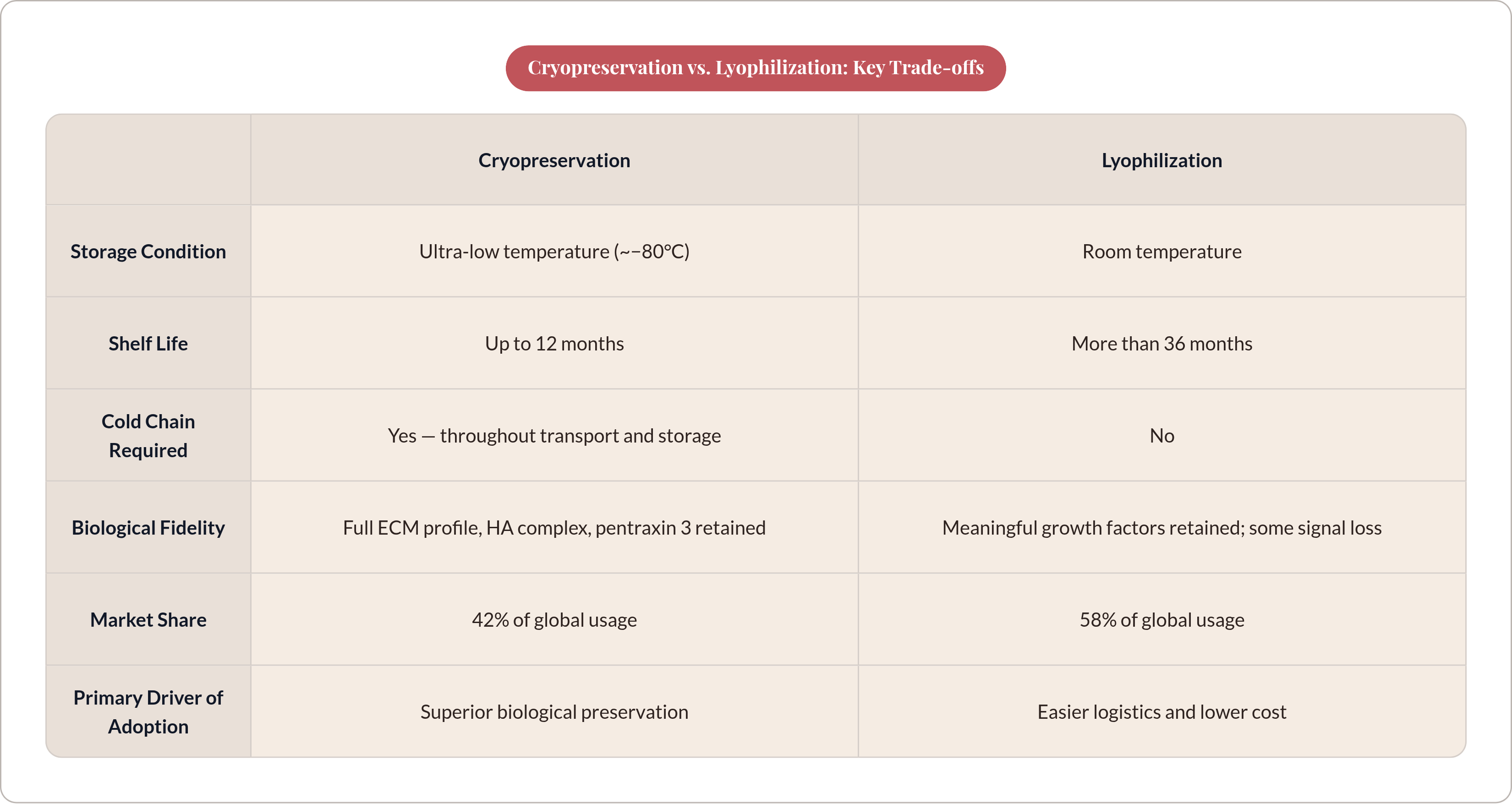
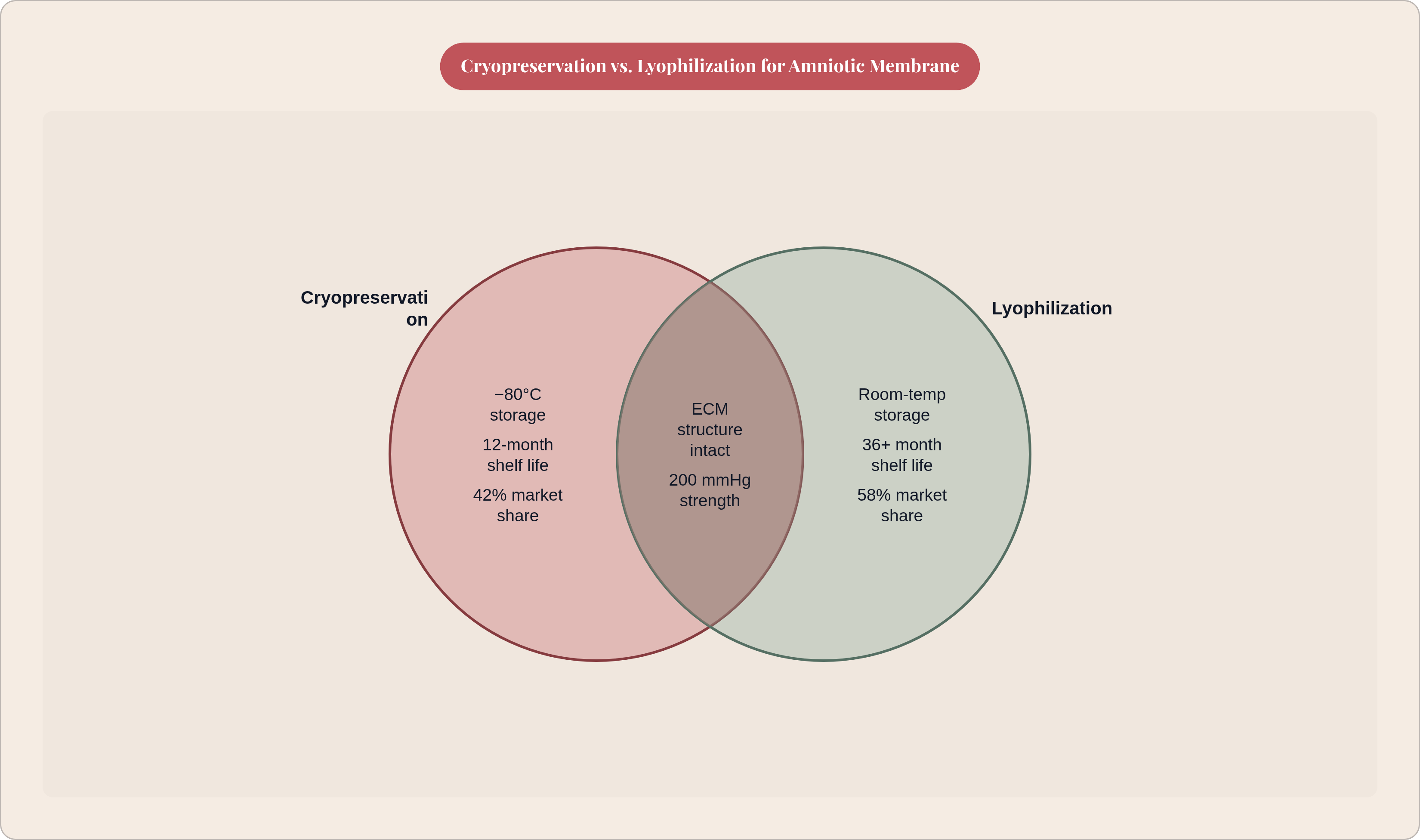
Cryopreservation stores the tissue at around minus 80°C in a glucose-based media, and it's the closest thing to freezing the biology in place. The native ECM architecture stays intact. High molecular weight hyaluronic acid, the heavy chain-HA complex, and pentraxin 3, a protein found in amniotic tissue, all stay present and active. Comparative research has found dehydrated tissues structurally worse off by comparison, almost entirely missing these components.
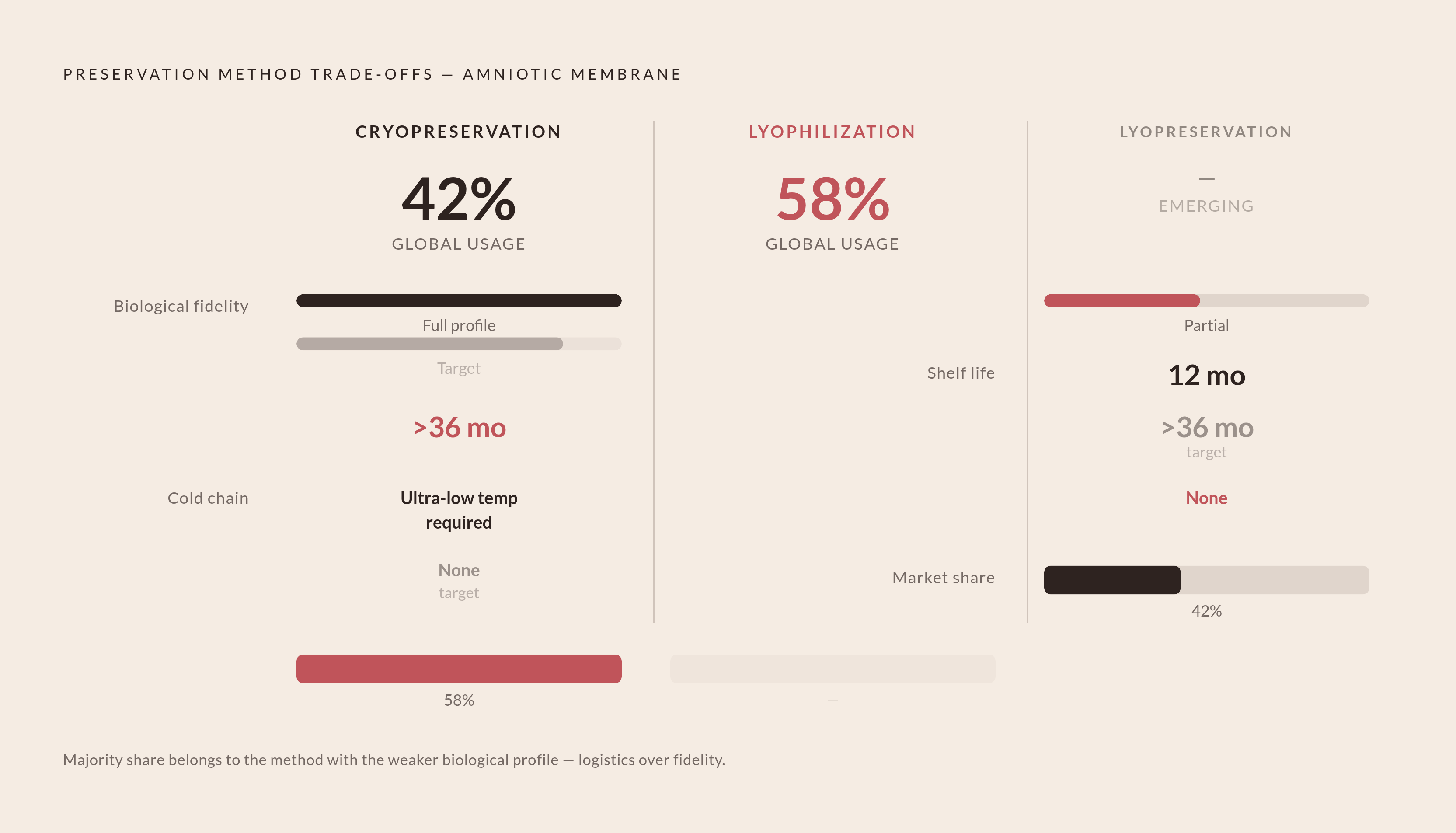
The catch is shelf life and logistics. Cryopreserved tissue is typically good for up to 12 months, and only if the cold chain never breaks, meaning ultra-low temperature freezers at the processing site, refrigerated transport the whole way, and ultra-low storage again at the clinic or surgical center. That's an expensive, fragile chain to maintain, and it shows up in the market: cryopreserved product makes up 42% of global usage, the minority share, despite having the stronger biological profile. That gap between what's biologically superior and what actually sells is worth sitting with, because it raises a real question. Are clinical outcomes tracking the biology, or are they tracking whatever's easiest to stock and ship? Lyophilization is the industry's answer to that logistics problem, and it comes with its own trade-off.
How lyophilization trades some biological fidelity for storage stability
Lyophilization is freeze-drying: pull the water out of the tissue, and it can sit at room temperature instead of needing a freezer. That single change solves most of the shipping and storage headaches that come with cryopreservation. It keeps the structural features and ECM architecture intact, along with a meaningful share of growth factors, but it doesn't hold onto the full biological signal profile the way cryopreservation does. Something gets left behind in the drying process.
What it buys back is time. Lyophilized product can sit on a shelf for more than 36 months, compared to roughly 12 months for cryopreserved alternatives, and it doesn't need a cold chain at any point. That difference alone explains why dehydrated product makes up 58% of global usage, the majority. A 2025 mechanical study published on ScienceDirect pressure-tested amniotic membranes preserved by cryopreservation, lyophilization, and dehydration, and all three held up to 200 mmHg without leaking or tearing. Structurally, the methods are close to equivalent. Biologically, they aren't.
So lyophilization's dominance in the market isn't a verdict on which method works better in a patient. It's a verdict on which method is cheaper to ship and easier to stock. Clinicians and procurement teams buying based on logistics should know that's what they're buying based on, not superior healing data.
There's a newer approach trying to split the difference: lyopreservation, a hybrid method aiming for cryopreservation's biological fidelity with lyophilization's room-temperature shelf life. Early research has shown tissue structure and cell viability after rehydration comparable to fresh amniotic membrane, though the data set is still young. BioTissue's CAM360 AmnioGraft, unveiled in December 2024, is a commercial example aimed at bypassing the cold-chain hurdles that come with conventional cryopreserved products.
How further processing transforms clean membrane into injectable and multi-layer formats
Historically, amniotic membrane was used as a flat sheet, laid over a wound or a damaged cornea. The injectable and multi-layer formats now on the market are a much more recent development, built on the same cleaned membrane but pushed through additional processing.
Micronization is one path. The tissue gets ground down, usually in a cryomill, under temperature-controlled conditions so the grinding heat doesn't cook off the bioactive content. Ground product targets a particle size under 1,000 micrometers; a minced alternative cuts the tissue into small cubes instead, somewhere between 0.1 mm and 3 mm. The output shows up as micronized dehydrated membrane, amniotic cytokine extract, or a solubilized powder meant for injection. These injectable forms are aimed at tendon, ligament, soft tissue, bone, and cartilage repair, and preclinical animal studies have shown they can promote healing in sports-related injuries, though this is still animal data, not settled clinical proof in humans.
The other path is building up instead of grinding down: laminated, multi-layer constructs that stack membrane sheets to add durability and concentrate biological activity for surgical implantation. More than 63% of new products introduced in 2025 used some form of multi-layer processing. Every format choice here, whether it's particle size, layer count, or how far the tissue gets solubilized, is the same preservation question asked in a different form: how much of the ECM's bioactive content survives being turned into this particular product.
How FDA classification shapes what a processor can claim and how a product reaches the market
Everything above eventually runs into a regulatory line, and where a product lands on that line depends on how it was processed, not just what it's made of. The governing framework sits in Sections 351 and 361 of the Public Health Service Act, with the actual mechanics spelled out in 21 CFR Part 1271.
Section 361 covers human cells, tissues, and cellular and tissue-based products, HCT/Ps in FDA shorthand, that meet certain criteria around how they are processed and used. Products qualifying under this pathway face a less burdensome regulatory path to market. Section 351 covers products that fall outside those criteria, and those are regulated as biologics, carrying a substantially more demanding regulatory burden before a product can be sold. The uncomfortable part is that the processing choices covered earlier in this piece, grinding, layering, solubilizing, are exactly the kinds of decisions that can push a product from 361 territory into 351 territory.
This isn't theoretical. In March 2024, MiMedx announced that the FDA had designated its AXIOFILL product a 351 biologic, and the company responded by initiating legal action contesting the ruling. That's a company with real regulatory and financial exposure fighting over exactly where the manufacturing line falls, which tells you how much is riding on that classification.
One thing doesn't change no matter which side of the line a product lands on: donor screening. The FDA requires screening for relevant communicable disease agents under 21 CFR Part 1271 Subpart C for nearly all HCT/Ps, with only limited statutory exemptions. So the processing method a company chooses is never just a technical or manufacturing decision. It's a regulatory one, and it determines two separate things at once: what the tissue is actually capable of doing in a patient, and what the manufacturer is legally allowed to say it does on the label.