Placenta-Derived MSCs vs Bone Marrow MSCs: Yield, Phenotype, and Expansion
Placental MSCs outperform bone marrow sources in yield and expansion capacity.

The placenta is not a single tissue. It contains both fetal and maternal compartments, and each yields MSC populations with meaningfully different biological properties. This is not a semantic distinction. When a published study reports results from "placenta MSCs," the specific sublayer under examination shapes everything that follows, and conflating those sublayers is one of the more durable sources of confusion in this field.
The major isolable regions include the amniotic epithelium, amniotic membrane, chorionic membrane, chorionic villi, chorionic trophoblast without villi, and the decidua, which is the maternal component. Work published in PLOS ONE in 2017 systematically characterized MSCs across these layers and found that chorionic membrane, chorionic trophoblast without villi, chorionic villi, and decidua-derived cells demonstrated better population doubling time and multilineage differentiation potential than cells from the amniotic epithelium and amniotic membrane. Decidua-derived cells showed the most favorable growth characteristics and the highest proliferation capacity relative to amniotic membrane and chorionic plate sources.
Amniotic membrane MSCs carry a different signature, expressing pluripotency markers OCT4, SSEA-3, and SSEA-4, alongside notably high secretion of pro-angiogenic factors including VEGF-A, angiopoietin-1, HGF, and FGF-2. That profile is not shared across the other layers. Chorionic villi MSCs, for their part, show elevated co-expression of CD146 and Stro-1, consistent with a pericyte-like subpopulation, and they demonstrate telomere maintenance properties with direct consequences for manufacturing runway.
Any rigorous comparison between placenta and bone marrow MSCs must specify which placental compartment is under examination. Studies that omit this detail are reporting on something. What that something is remains unclear.
The Yield Gap — What a Single Placenta Can Produce Versus What Bone Marrow Requires
MSCs are rare in bone marrow, and they become rarer in elderly donors as the frequency of MSC progenitors within the stromal compartment declines measurably with age. This is a structural problem that process optimization can't resolve. Generating clinically meaningful cell numbers from bone marrow requires extensive ex vivo expansion, and expansion is precisely where bone marrow MSCs begin to lose ground. Potency decreases with passage number, and the more expansion required to reach a therapeutic dose, the more degraded the product at journey's end.
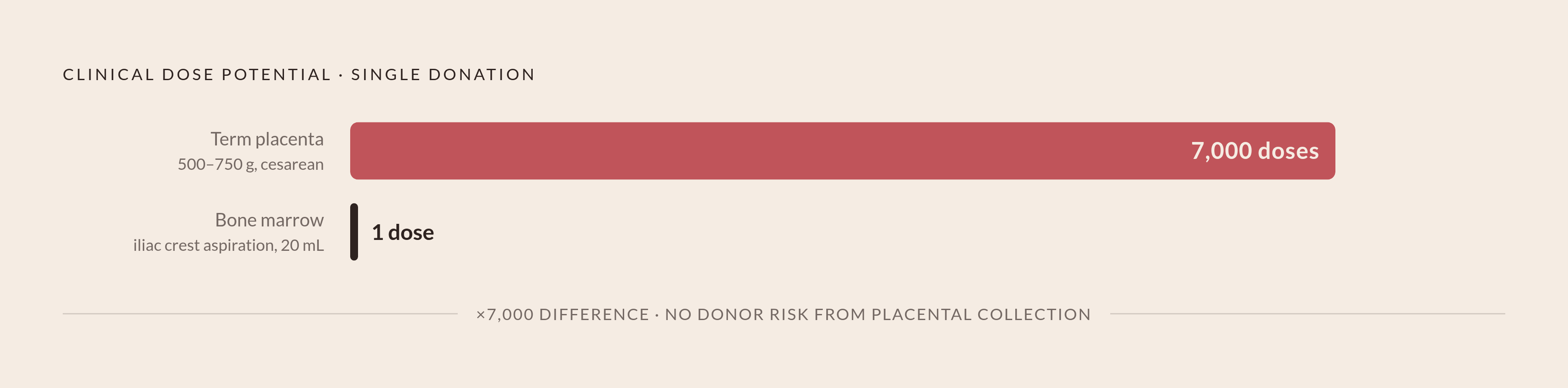
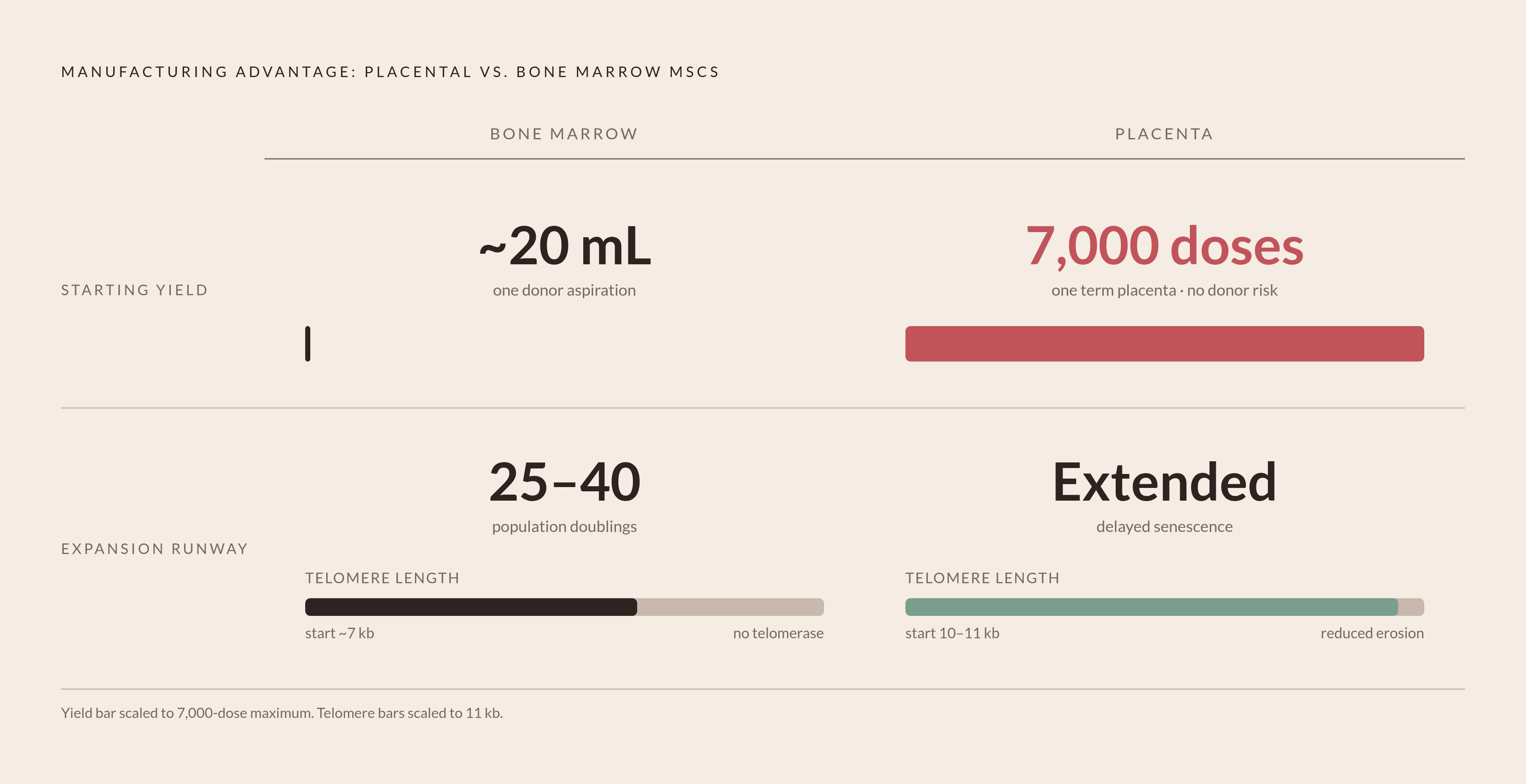
Bone marrow collection by iliac crest aspiration yields approximately 20 mL per procedure from a volunteer donor. The donor bears real medical risk. The starting volume is fixed and small.
A term placenta weighs between 500 and 750 grams, is collected aseptically during cesarean section, carries no donor risk, and is otherwise discarded. A single term placenta has been demonstrated to contain sufficient MSC progenitor material to manufacture up to 7,000 clinical doses. That scale difference is not incremental. It is categorical.
Under GMP conditions, a 2025 Scientific Reports study measured this directly. Umbilical cord tissue yielded 6.5 × 10⁶ ± 0.8 viable MSCs per sample; amniotic fluid yielded 5.8 × 10⁶ under identical conditions. Tissue type was the strongest predictor of MSC yield, with a standardized beta coefficient of 0.61. Source selection is the primary lever in yield optimization. Process variation is secondary.
That same study reported something that deserves to be stated plainly rather than buried: placental tissues showed microbial contamination rates of 18 to 21 percent under GMP isolation conditions, compared to 3 percent for amniotic fluid. Programs that subordinate sterility to productivity are where failures originate. This isn't a footnote; it's a process design constraint that demands the same rigor as any yield target.
The age-related decline in BM-MSC proliferation compounds the problem. Older donors produce fewer starting cells, those cells expand more slowly, and they senesce earlier. These constraints are upstream of anything a manufacturing process engineer can address.
Where Placenta MSCs and BM-MSCs Look the Same — The Shared ISCT Surface Marker Profile
In 2006, the International Society for Cell and Gene Therapy established minimal criteria for defining human MSCs, requiring positive expression of CD73, CD90, and CD105, alongside absence of hematopoietic markers CD14, CD19, CD34, CD45, and HLA-DR. These criteria became the definitional floor for MSC identity regardless of tissue source.
CD73, CD90, and CD105 are not true stemness epitopes. Their selection reflected practical consensus, not mechanistic insight. They define a category, not a function. That said, they matter enormously for regulatory and clinical comparability arguments, because allogeneic placenta-derived MSCs can be positioned against the same identity criteria applied to established bone marrow products. The shared marker profile is the regulatory bridge between sources. Whether that bridge adequately captures biological differences between those sources is a question the field has been wrestling with since the criteria were published.
Across Wharton's jelly, adipose tissue, bone marrow, and placental MSCs, expression of CD26, CD34, CD44, CD73, CD90, CD166, and HLA-ABC is broadly similar. Placenta-derived and umbilical cord MSCs also express low or undetectable levels of class II HLA, consistent with BM-MSCs. This low alloimmunogenicity profile is foundational to the case for HLA-unmatched, off-the-shelf allogeneic use.
The convergence at ISCT markers is a shared entry point, nothing more. The biology that actually distinguishes these sources begins where this common floor ends.
Where Phenotype Diverges — Integrin Expression, Differentiation Lineages, and Immunomodulatory Potency
Beyond the ISCT floor, significant inter-source differences emerge across marker classes including CD14, CD56, CD79a, CD105, CD106, CD146, and CD271. These are not minor variations in expression intensity. They reflect genuinely different biology, with functional consequences in both therapeutic and manufacturing contexts.
Integrin expression is one area where divergence carries direct practical weight. Placenta-derived MSCs attach well to fibronectin-derived peptides P7, P15, and P17. BM-MSCs bind P7 but show minimal attachment to P15 and P17, driven by distinct expression profiles across integrin alpha-2, alpha-4, and alpha-6 chains. For scaffold-based tissue engineering applications, this distinction enters the equation at biomaterial selection, not as an afterthought.
Differentiation lineage differences follow a consistent pattern. Placenta-derived MSCs are reliably less capable of adipogenic differentiation than BM-MSCs. On the myogenic side, the TGF-beta-1, PDGF, and ascorbic acid protocol sufficient to produce smooth muscle-like cells from BM-MSCs fails entirely in placenta MSCs; a distinct protocol is required. Osteogenic differentiation shows more granularity: amniotic membrane and umbilical cord MSCs differentiate toward the osteoblast stage more efficiently and more rapidly than chorionic membrane and decidua MSCs, with fibronectin augmenting this process through Akt and ERK phosphorylation.
Immunomodulatory profile is where the comparison becomes most consequential for therapeutic positioning. Placenta-derived MSCs affect antigen-presenting function in mononuclear cells and dendritic cells more significantly than cord-derived MSCs. Their conditioned medium produces measurable reduction in T cell proliferation relative to cord-MSC conditioned medium. Co-culture increases IL-10 and TGF-beta-1 while reducing IFN-gamma, and it specifically enriches CD3+CD4+CD25+ T regulatory cells, supported by increased FoxP3 mRNA expression. That regulatory T cell induction capacity appears mechanistically distinct from what BM-MSCs produce.
The complication: in PHA-stimulated assay conditions, placenta-derived MSCs show only weak inhibition of T cell proliferation, whereas BM-MSCs produce a clear and measurable reduction. BM-MSCs retain a genuine immunosuppressive advantage in specific stimulation contexts. Any characterization that glosses over this is claiming more than the data supports.
Expansion Capacity and Why Telomere Biology Gives Placenta-Derived MSCs a Manufacturing Runway BM Cannot Match
Every primary MSC population undergoes replicative senescence. Telomeres shorten with each division, and when they reach a critical threshold length, senescence-signaling pathways activate and proliferation ceases. In a manufacturing process, that's a hard ceiling on the number of doses derivable from any starting population. It can't be engineered away.
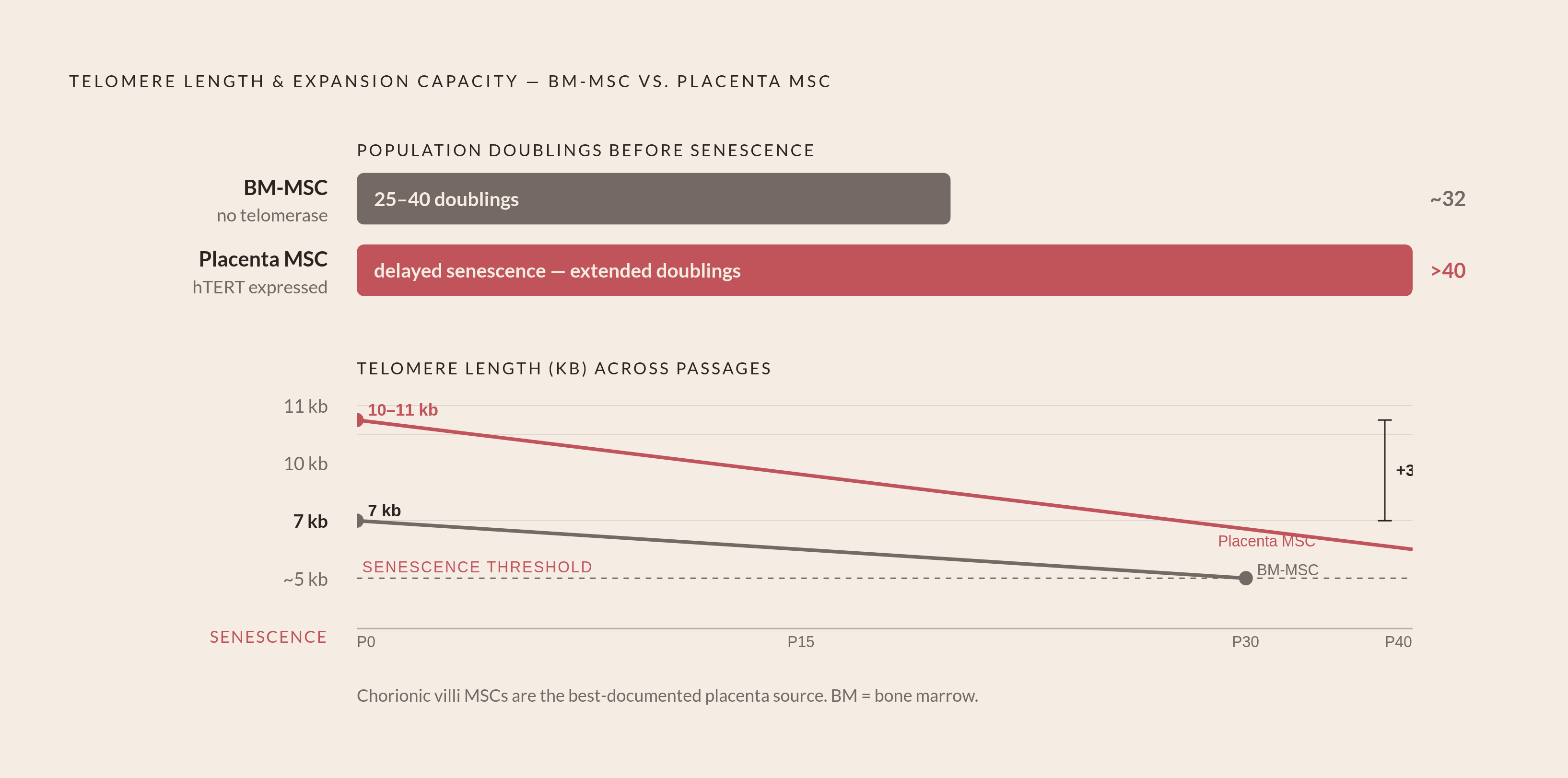
BM-MSCs undergo roughly 25 to 40 population doublings before senescence. No telomerase activity has been detected under standard conditions, and telomere length decreases with passage in a well-documented, linear fashion. The ceiling arrives early in the expansion process, at the moment manufacturing demands the most headroom.
Telomere length at early passage is strongly donor-age-dependent. Fetal tissue MSCs begin with telomere lengths in the range of 10 to 11 kilobases; postnatal sources start at approximately 7 kilobases. Because placenta is perinatal tissue, its MSCs enter culture with a longer starting runway. They reach therapeutic dose targets in fewer passages and accumulate less expansion-induced potency drift along the way.
Chorionic villi MSCs are the best-documented case. They demonstrate reduced telomere erosion and delayed senescence relative to MSCs from at least three other tissue sources. Increased hTERT gene expression has been observed, though researchers have noted that hTERT expression alone can't fully account for the maintenance observed; the underlying mechanism remains unresolved. What is not unresolved is the functional outcome: CV-MSCs retain multilineage differentiation potential and extracellular matrix remodeling properties through extended culture. The expansion advantage does not hollow out the cells in the process of exploiting it.
There is also a maternal donor age effect with direct banking implications. A 2021 study in Aging found that self-renewal capacity was highest in donors aged 26 to 30, that expression of NANOG, OCT4, and SSEA4 was higher in donors aged 22 to 35, and that adipogenic differentiation increased up to sevenfold within that window. Donor selection criteria are not incidental to placental MSC banking strategy. They're determinative of product quality, and treating them as administrative detail is a costly mistake.
On genomic stability, SNP GeneChip analysis has confirmed that no copy number variations are acquired during sequential passaging in either placenta or BM-MSCs. For longer expansion runs, which placental manufacturing requires, this is a foundational GMP safety finding. It should be treated as such.
What the Manufacturing Case for Allogeneic Placenta MSCs Looks Like When Yield, Phenotype, and Expansion Are Read Together
The placenta is not a theoretically interesting alternative to bone marrow. It's a materially different starting point, and the differences compound across the manufacturing process in ways that consistently favor it for allogeneic, multi-dose production. A large starting yield reduces expansion burden. A longer replicative lifespan means fewer passages to reach a therapeutic dose. Low class II HLA expression supports HLA-unmatched, off-the-shelf positioning grounded in measurable biology. Each of these properties amplifies the others.
The infrastructure to act on this is in place. GMP-compliant production protocols for placental MSCs have been described in a 2024 chapter of Methods in Molecular Biology. A 2025 Scientific Reports phase 1 trial used GMP-manufactured placenta-derived MSCs in secondary progressive multiple sclerosis. The manufacturing rationale is already being stress-tested in early clinical settings, which is the only test that ultimately matters. Most platform arguments for novel cell sources collapse somewhere between bench characterization and first-in-human data. This one has not.
The unresolved problems deserve the same candor. Microbial contamination rates of 18 to 21 percent under GMP isolation conditions remain a process engineering challenge without a fully satisfactory solution. Robust long-term immunological safety data from allogeneic MSC trials are limited across all sources, including bone marrow. Scaling from academic GMP batch production to volumes sufficient for a phase III trial requires industrial infrastructure that hasn't yet been demonstrated at full commercial scale for this source.
Bone marrow's clinical precedence is real and was earned through decades of rigorous work. But clinical precedence doesn't resolve a manufacturing problem that is biological in origin. The starting material sets the ceiling on what any process can achieve. For allogeneic, multi-dose production, the placenta sets that ceiling higher.