Placental Growth Factors and Their Therapeutic Applications
Scientists map how placental growth factor blocks tumors and predicts pregnancy complications.

Persico and colleagues discovered placental growth factor in 1991 and described it as an angiogenic factor and member of the VEGF family. The broader tissue expression of PlGF has since become clear. The protein runs 149 amino acids, folds into a dimer, and carries N-linked sugar chains, and crystallography has mapped its structure down to 2.0 Å, so we know exactly how it folds and where it grips its receptor.
It shares about 53% of its amino acid sequence with VEGF, close enough to use the same receptor machinery, distinct enough to produce effects VEGF doesn't.
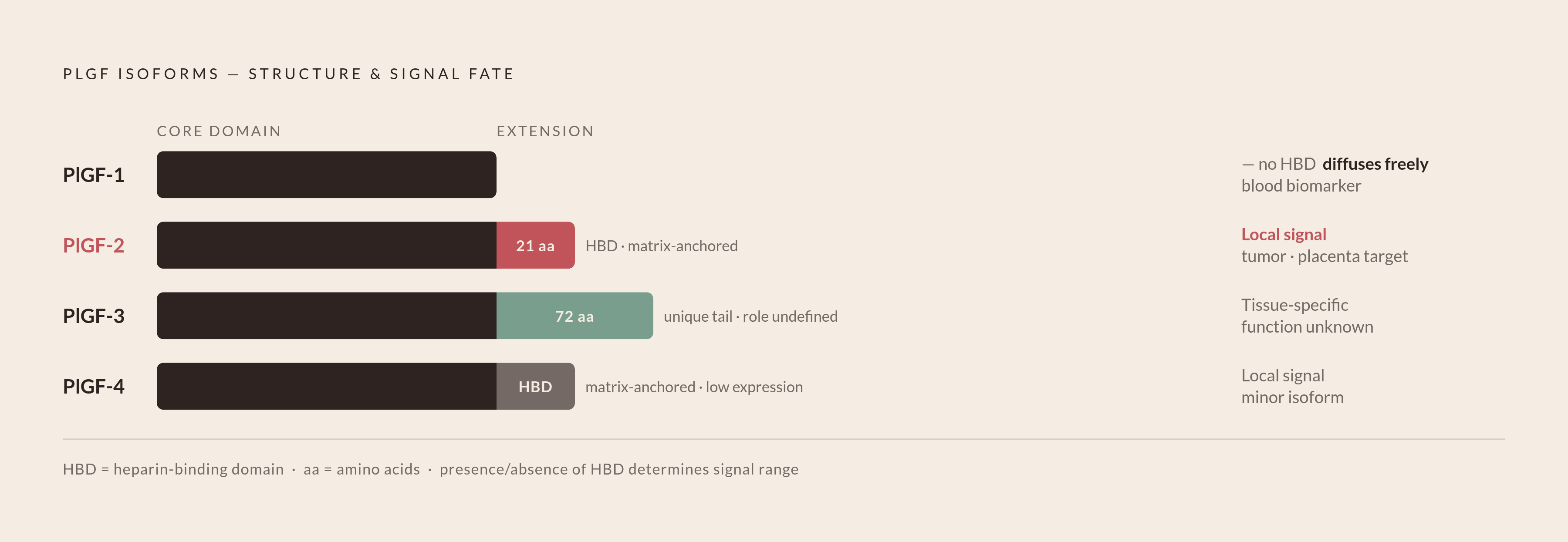
Four splice isoforms exist, and where each one ends up in the body changes what it does there. PlGF-1 has no heparin-binding domain, so it drifts freely through blood and tissue rather than anchoring to the extracellular matrix, while PlGF-2 carries a 21-amino-acid heparin-binding domain that anchors it to the extracellular matrix and to cell surfaces, keeping its signal local instead of systemic, and this localized signaling is central to its roles in tumors and in the placenta. PlGF-3 carries a unique 72-amino-acid tail on its C-terminus, and nobody has worked out what that tail does in specific tissues yet. PlGF-4 runs longest of the four and binds the matrix the way PlGF-2 does, but shows up at lower levels wherever it's found.
That heparin-binding domain sounds small, yet it governs a great deal. Any isoform carrying it, PlGF-2 especially, grabs onto heparan sulfate chains on cell surfaces the same way VEGF165a does, and whether an isoform stays put inside a tumor or a placenta, or travels through blood where a lab can draw and measure it, comes down to that one feature. Oncology leans on it, and so does screening for poor blood flow in pregnancy.
PlGF turns up well past the placenta too, showing up in heart tissue, lungs, thyroid, fat, and skeletal muscle. That spread is why its reach runs into cardiology and oncology, not just obstetrics.
How PlGF's receptor signaling produces disease-selective angiogenesis
PlGF binds mainly to VEGFR-1, also called Flt-1, and only reaches VEGFR-2 (Flk-1) indirectly, through crosstalk between the two receptors. When PlGF activates VEGFR-1, the phosphorylation pattern differs from what VEGF triggers there, and it runs its own signal, with its own downstream consequences.
VEGF-A and PlGF can also pair up into a heterodimer, and that combined molecule binds VEGFR-1 with a stronger preference than either protein manages alone. In tumor vessels or ischemic tissue, that heterodimer effect carries real weight.
Here's the property that makes PlGF worth targeting in the first place. Knock it out, or block it with a drug, and the quiet, already-formed vessels in healthy tissue stay untouched, while only new vessel growth in disease states gets cut off. Anti-PlGF treatment stops tumor growth even in tumors that have already gone resistant to VEGF-targeted drugs, and it does this without damaging normal blood vessels. VEGF monotherapy has never managed that safety profile.
Preclinical work backs a second point. Block VEGF and PlGF at the same time, or hit VEGFR-2 and VEGFR-1 together, and tumor angiogenesis drops harder than either block alone manages. Every dual-target drug in oncology and ophthalmology traces back to that one finding.
PlGF as a biomarker for preeclampsia: from pathophysiology to FDA-cleared testing
Preeclampsia complicates a meaningful share of pregnancies worldwide and drives a large chunk of maternal and neonatal illness. Clinicians have wanted a reliable early-warning test for decades.
The biology gives them a clean signal to work with. In a normal pregnancy, PlGF in the mother's blood climbs steadily through gestation and peaks around 30 weeks, while in preeclampsia, it drops below that curve, and it drops before symptoms show up. A low reading can flag risk before a patient develops high blood pressure or protein in her urine.
Pairing PlGF with sFlt-1, an antiangiogenic protein that works against it, sharpens the picture further. The ratio between the two, sFlt-1 over PlGF, reliably predicts which patients go on to develop preeclampsia with severe features.
In 2023, the FDA cleared a serum sFlt-1/PlGF test with a cutoff of 40, meant to assess risk in hospitalized women with hypertensive disorders of pregnancy between 23 and 35 weeks. The clearance marked a significant regulatory milestone, and assay platforms for this test now run in hospital laboratories.
The performance numbers hold up under scrutiny. In the PELICAN study, a PlGF reading at or above 100 pg/ml ruled out delivery-required preeclampsia within 14 days, with a negative predictive value of 0.98, while in the PROGNOSIS study, an sFlt-1/PlGF ratio at or below 38 ruled out delivery within one week, with a negative predictive value of 99.3%. Neither number tells a clinician who will get sick, but both tell her who almost certainly won't get sick soon, and on a labor and delivery unit, that's the number she actually needs. A negative result means extending monitoring safely instead of rushing toward intervention.
The PARROT-2 trial, published in The Lancet in February 2024, gave the strongest evidence yet that this testing changes outcomes, beyond diagnostic accuracy alone. The multicenter randomized trial found that PlGF-based testing shortened time to diagnosis and cut severe maternal complications. PARROT-2 demonstrated that the test's diagnostic accuracy translated into measurable improvements in patient care.
Term preeclampsia is still unsolved. The sFlt-1/PlGF ratio measured at 35 to 37 weeks predicts it with roughly 80% detection, and the PE37 study, published in BMJ Open in March 2024, is testing whether screening-guided delivery timing at that window stops cases from progressing. Results from that trial will help determine whether the question can be closed.
Newer modeling from 2025 combines PlGF with uterine artery pulsatility index and mean arterial pressure, measured in the first trimester. The field is early, but multi-parameter prediction is gaining ground over PlGF measured alone. A 2025 review from researchers at Mount Sinai and the University of Toronto described PlGF and sFlt-1 as tools that now guide routine obstetric and medical care in pregnancy, extending well beyond specialist referral.
Targeting PlGF in cancer: aflibercept and the case for dual VEGF/PlGF blockade
Tumors make their own PlGF, and so does the stromal tissue around them. Across solid tumor types, higher PlGF levels track with more advanced stage, greater odds of metastasis, and worse survival, a relationship that has been documented in the research literature.
Aflibercept, marketed as a VEGF Trap, fuses the extracellular binding domains of VEGFR-1 and VEGFR-2 into one decoy receptor. That decoy soaks up VEGF-A, VEGF-B, and both PlGF-1 and PlGF-2 before they reach their real receptors on tumor blood vessels, cutting angiogenic signaling off through several channels at once.
The Phase III VELOUR trial tested aflibercept plus FOLFIRI chemotherapy against placebo plus FOLFIRI in patients with metastatic colorectal cancer who had already progressed on oxaliplatin. The combination produced statistically significant gains in both progression-free survival and overall survival, which led to FDA approval for this specific indication. Grade 3 and 4 adverse events showed up more often in the aflibercept arm, though, and that toxicity tradeoff is one any clinician prescribing it has to weigh, not something to skim past on the label.
The mechanism explains why this beats single-target VEGF drugs. Blocking VEGF and PlGF together suppresses tumor angiogenesis synergistically in preclinical models, and neither target alone gets you there. PlGF blockade carries its own specific advantage too: it stops tumor growth in cancers already resistant to VEGFR inhibitors, without touching normal vasculature, which is exactly the failure mode anti-VEGF monotherapy keeps running into.
The patent filings track the momentum. Over 120 active patents filed between 2024 and 2026 cover new delivery systems, combination regimens, and biomarker applications tied to PlGF. Most of these compounds haven't proven anything in humans yet, but the volume of filings says something about where the field expects things to go.
What hasn't happened yet is the head-to-head Phase III trial that would settle the question. The preclinical case for anti-PlGF strategies beating anti-VEGF monotherapy is strong, but the regulatory proof still doesn't exist.
PlGF inhibition in retinal disease and where multi-target ophthalmology is heading
The scale of retinal disease alone justifies the investment. An estimated 18.8 million adults worldwide had clinically significant diabetic macular edema in 2020, and that number is set to grow substantially by 2045.
Aflibercept shows up again here, reformulated for injection directly into the eye and sold as Eylea and Eylea HD. It uses the same trap mechanism as the oncology version, binding VEGF-A, VEGF-B, and PlGF, and it's approved for neovascular age-related macular degeneration, diabetic macular edema, and diabetic retinopathy.
The American Academy of Ophthalmology updated its guidelines in 2024, reaffirming intravitreal anti-VEGF treatment as first-line therapy for AMD and diabetic eye disease, with more emphasis than before on agents that hit multiple targets. Co-targeting PlGF alongside VEGF has gained increasing emphasis, reflected in the updated guidelines.
Faricimab took a related but different path. It's the first bispecific antibody approved for intravitreal injection, targeting VEGF-A and angiopoietin-2 rather than PlGF directly, and it performed as well as aflibercept on visual acuity and central macular thickness in trials. PlGF isn't on its target list, yet the result backs the same underlying logic: two vascular pathways beat one, and that principle reaches past the VEGF/PlGF pairing specifically.
The next drugs in the pipeline aim wider still, going after VEGF-C, VEGF-D, and their receptors alongside PlGF, targeting vascular inflammation and lymphatic vessel growth that single-target drugs miss. Ophthalmology has accumulated substantial clinical experience with PlGF-inclusive drugs, and that experience reflects how combination targeting has matured in this field.
PlGF's emerging role in cardiovascular repair and ischemic disease
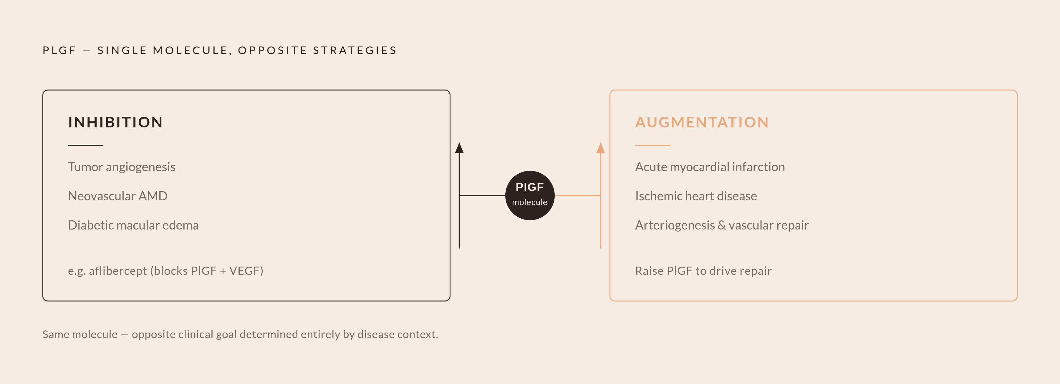
Cardiology flips the whole logic around. In the tumor and eye, the goal is blocking PlGF, while in ischemic heart tissue, PlGF does repair work, so the goal becomes raising it instead.
PlGF switches on under cellular stress and drives vascular repair once it's expressed. In patients recovering from acute myocardial infarction, the level of PlGF expressed in cardiac tissue predicts how much left ventricular function comes back.
Preclinical gene therapy work, delivering the PlGF gene directly into heart tissue after a heart attack, improves cardiac performance through several mechanisms at once. It triggers new blood vessel growth in the heart muscle, boosts paracrine signals that block cell death and encourage angiogenesis, and pushes bone marrow to produce more progenitor cells and release them into circulation. It also nudges bone marrow cells to switch identity toward endothelial cells, smooth muscle cells, and even cardiomyocytes.
The net effect in these models is a smaller infarct area and better cardiac function overall, driven both by new capillary growth and by the enlargement of existing arteries, a process called arteriogenesis. That's a different mechanism from capillary sprouting, and it may matter more for restoring real blood flow to starved tissue.
There's a metabolic angle here too. A 2024 study by Park and colleagues, published in Metabolism, found that PlGF deficiency triggers metabolic syndrome tied to obesity and aging. That pushes PlGF's relevance past acute heart attacks and into chronic disease prevention, a slower and much larger target than a single infarct.
None of this sits close to the clinic, since cardiovascular PlGF therapy remains in preclinical and early translational stages. The biology looks promising, but no clear clinical timeline has emerged.
How PlGF's isoform diversity and disease selectivity shape the next generation of therapeutics
The isoform differences from earlier aren't a footnote; they explain why PlGF ends up in such different clinical tools. PlGF-2's matrix-anchoring domain keeps its signal concentrated inside tumors and the placenta, which is exactly why it matters so much in oncology, while PlGF-1's freedom to diffuse through blood is what makes it the biomarker of choice in preeclampsia screening. Same protein family, same receptor biology, but which isoform is in play decides whether a lab ends up building a blood test or a drug company ends up building a cancer drug.
Disease selectivity runs under all of it. PlGF stays mostly idle in healthy tissue and switches on during disease, so drugs built around it carry a narrower risk of hurting healthy tissue than broad VEGF blockade does. That difference is the argument for keeping PlGF-specific research funded instead of settling for VEGF drugs alone.
Combination targeting keeps showing up: the sFlt-1/PlGF ratio in obstetrics, dual VEGF and PlGF blockade in oncology and retinal disease, PlGF paired with uterine artery pulsatility index and mean arterial pressure in first-trimester screening. PlGF does its best work as one signal inside a larger system.
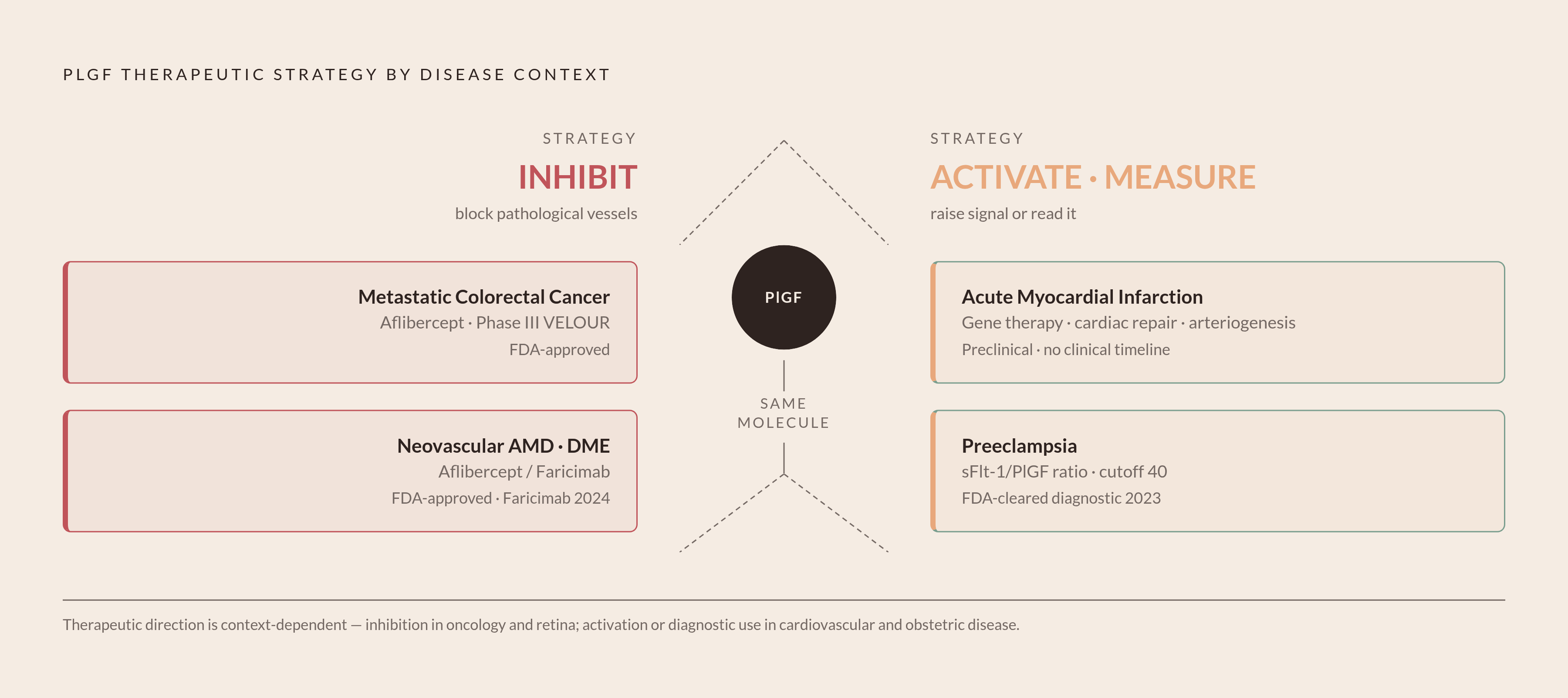
The therapeutic strategy splits clean along disease lines. Where PlGF drives pathological vessel growth, in cancer and retinal disease, the goal is inhibition, while where it drives tissue repair, in ischemia and cardiac recovery, the goal is raising it back up. Same molecule, and which intervention a physician reaches for depends entirely on which side of the disease she's standing on.
Real gaps remain, and they're worth naming plainly instead of smoothing over. PlGF-3's tissue-specific role is still undefined, and whatever isoform-selective drugs might come from understanding it haven't been mapped out. Anti-PlGF-specific agents, as opposed to dual VEGF/PlGF blockers like aflibercept, haven't reached pivotal Phase III trials in most disease areas. And getting cardiovascular gene therapy out of animal models and into humans remains the biggest open question in that corner of the field, one that won't close soon.
PlGF went from a pregnancy-specific curiosity, described by Persico's team in 1991, to a molecule with FDA-cleared diagnostic and therapeutic uses across obstetrics, oncology, and ophthalmology. It took thirty-some years of structural biology, disease-selective pharmacology, and a fair amount of unmet clinical need to get there, and the cardiology chapter hasn't even started yet.