Placenta Stem Cell Harvesting Techniques and Yield Factors
Different harvesting methods yield different cell populations, each with tradeoffs.

The placenta carries three distinct, clinically useful cell populations in one organ: hematopoietic stem cells (HSCs), mesenchymal stem cells (MSCs), and, for newer oncology work, placental circulating T (P-T) cells, which are immune cells rather than a stem cell type. Yield from this organ depends on a predictable set of biological, procedural, and timing variables, and the technique chosen to harvest it determines which of those three populations a given program can actually use.
The placenta as a productive but structurally complex harvest source
The placenta is not one tissue but several, stitched together into a single discarded organ at the moment of birth. Each of its three major cell populations sits in its own compartment and calls for its own isolation approach, so the organ's richness and its difficulty arrive together, as the same structural fact. That the tissue would otherwise be thrown away, and that collection adds no invasive step beyond delivery itself, makes the ethical case for harvesting it about as clean as these cases get in regenerative medicine. The immunology adds a practical reason to care: placental MSCs carry low or absent expression of human leukocyte antigens class II and other co-stimulatory molecules, along with intrinsic immunomodulatory properties, which lets them be transplanted into unrelated recipients without the matching requirements that burden most allogeneic cell therapy. HSCs drawn from placenta and umbilical cord blood are already established as a proliferative, high-value alternative to bone marrow and peripheral blood collection. None of this changes the underlying problem: chorionic villi, amniotic membrane, decidua, the placental vasculature, and the cord are different tissue types with different cell populations embedded in them, and no single harvesting method reaches all of them with equal efficiency. Choosing how to harvest the placenta is, in practice, choosing which of its populations to prioritize, and that choice shapes everything downstream of it.
The four principal isolation methods
Each solves a different piece of the harvest problem, and each gives something up to do it, so that none of the four wins outright on every measure that matters.
Vascular perfusion works by flushing a protease solution through the placental blood vessels, producing a cell population that is almost entirely fetal in origin. Some reports credit it with higher raw cell counts, but when evaluated specifically for chorionic villi MSCs, perfusion showed the lowest efficiency for isolation and uniformity of any method tested, a gap between the volume it produces and the volume actually usable afterward. Keeping a vascular access route sterile at clinical scale also adds logistical weight that the other three methods avoid.
Enzymatic digestion is the clinical workhorse, the method most labs reach for first. It breaks tissue apart mechanically using proteases, commonly a combination of Trypsin and Collagenase. That combination produces a meaningfully higher cell count than Trypsin used alone, a direct result of more thorough tissue breakdown. The trade-off is biological: extended exposure to the enzymes lowers viability and can change MSC proliferative capacity, so the method that yields the most cells on paper can yield a batch that performs worse in culture. A 2025 protocol from Huasan Biotech tried to close that gap from the production side rather than the biology side, running the digestion in sterile, closed blender bags with collagenase I at 100 U/mL, dispase at 2.5 U/mL, and DNase I at 100 U/mL, built specifically to cut the safety risk and labor cost of manual open processing at commercial volume.
Explant culture skips enzymes. Tissue fragments go straight into culture, and MSCs migrate out of them on their own over roughly 14 days. That slower path avoids the viability penalty that enzymatic exposure imposes, and the cells that do emerge tend to show better viability and quality, but a two-week turnaround is hard to reconcile with a clinical timeline under any pressure.
Modified explant culture tries to close that gap directly: a brief, mild enzymatic step speeds up the early phase, followed by standard explant culture to protect cell quality through the rest of the process. It was built specifically to answer the speed problem that pure explant culture has, while still meeting the volume and quality bar that clinical trials require. What it really changes is the nature of the choice: instead of picking speed or quality, modified explant culture lets a lab dial the balance between them. That dial, more than any single method's raw output, is the real axis these four techniques compete on.
Collection timing, delivery mode, and the ceiling on technique performance
Before any laboratory technique touches the tissue, two decisions made in the delivery room already set limits on what that technique can achieve: when the placenta comes out, and how.
Delivery mode shapes contamination risk directly. Placental fragments and whole placenta carry the highest contamination levels among perinatal tissues, a result tied to prolonged exposure to the birth canal and maternal blood during vaginal delivery. A Scientific Reports study led by Çopuroğlu and colleagues analyzed vaginally delivered samples as a separate group specifically to confirm this elevated contamination tendency. Caesarean delivery is favored for GMP-grade placental harvest for this reason, a contamination-control decision with direct bearing on whether the resulting tissue meets clinical-grade standards, not a matter of institutional habit.
Timing after delivery carries its own cost. Established protocols treat harvesting between roughly zero and two hours post-expulsion as a workable window. Some protocols allow sterile storage at 5 to 25°C for an extended period, but the better practice keeps the exsanguinated placenta stored no more than 36 hours before stem cell collection begins. Every hour past the optimal window adds cell death and contamination risk that no technique applied afterward can fully reverse. The operational consequence is straightforward: a biobank or clinical program has to manage delivery-room logistics, coordinating with obstetric staff on delivery mode and transport timing, with the same rigor it applies to the laboratory protocol itself. A perfect isolation method run on compromised starting material still produces a compromised yield.
Biological variables in the donor and tissue that shift yield within a given method
Once delivery mode and timing are controlled, the remaining variation in yield traces back to the donor and to which part of the placenta a protocol actually draws from.
Tissue compartment turns out to be the single strongest predictor of viable MSC yield. The Çopuroğlu et al. Scientific Reports study, the first head-to-head GMP-conditions comparison of all four perinatal sources, found tissue type to be the strongest predictor of viable MSC yield. Umbilical cord-derived MSCs produced the highest mean yield per sample, amniotic fluid and amniotic membrane followed, and whole placenta and placental fragments trailed with significantly fewer viable MSCs under the same conditions. "Placental stem cells" functions less as one category than as a set of sources with real differences in productive potential.
Donor characteristics add a second layer. A 2024 Scientific Reports finding tied higher maternal age strongly to longer cell doubling time, so cells from older donors expand more slowly in culture, and a batch can look adequate at the point of isolation while still underperforming later for exactly this reason. Gestational diabetes carries a similar penalty: compared to normal samples, the proliferation rate of early-passage GDM samples is significantly lower, an underappreciated screening criterion for biobank procurement. Other obstetric details that intuitively seem relevant, gestational age, birth weight, and cord breadth, were not found to independently predict Wharton's Jelly MSC yield, which suggests maternal metabolic history is a better forecasting tool than the routine delivery-room chart.
A separate problem concerns where the cells actually come from, biologically rather than anatomically. Karyotype analysis has shown that decidua-derived MSCs are maternal in origin, while the placenta's other cell types trace to the fetus, so a protocol that processes whole placenta without dissecting it by compartment first can end up with a batch that mixes maternal and fetal cells without anyone realizing it. This is not a minor technical footnote. Some culture methods have produced MSCs of maternal origin even when the anatomical starting material was fetal chorionic villi, an identity mismatch that complicates reproducibility across labs and raises real questions for any regulator trying to confirm what a given cell product actually is. Knowing which donor and tissue variables drive yield only pays off if the collection method is built to capture enough of the right cells in the first place, which is exactly where the next population, HSCs, runs into trouble.
Pulsatile perfusion and the HSC volume constraint
For HSCs, the limiting factor isn't the sophistication of any single technique but sheer volume. Current collection methods leave a large share of individual cord blood and placental collections too small to use on their own, a shortfall that affects even pediatric patients who need comparatively fewer cells per treatment. This is a structural constraint, not a technique failure: the biology of HSC distribution across the placenta and cord does not concentrate enough cells at any single access point when collection relies on conventional needle-and-syringe withdrawal or passive gravity drainage.
Pulsatile perfusion offers a partial answer. Running the residual placental cord blood through machine-driven pulsatile perfusion produces a cell population more enriched for primitive hematopoietic stem cell markers, specifically CD133+, than conventional venipuncture withdrawal manages. The mechanism is pressure cycling through the vasculature in controlled pulses, which reaches HSCs sitting in deeper tissue compartments that simple passive drainage never disturbs. It's in the proportion of that collection made up of primitive, engraftment-capable HSCs, which determines engraftment success in clinical transplantation independent of the raw cell count on the label.
Celularity's CAR-T work and optimized placental T-cell harvest
Placental T cells have drawn far less banking and research attention than HSCs or MSCs, yet they're now showing clinical value tied directly to their placental origin, properties that T cells from conventional donor sources don't carry. Celularity built a defined process for generating and expanding CD19 CAR-T cells from human placenta, and the resulting cells show strong anti-tumor activity in both lab and animal models with little sign of triggering acute graft-versus-host disease, the complication that normally makes donor T cells hard to use across unrelated recipients. In mid-2024, Celularity moved a related program forward, announcing the start of a Phase I trial for an allogeneic NK cell therapy derived from placental cells, aimed at multiple myeloma, carrying this collection and expansion process into a regulated clinical setting.
The appeal of an "off-the-shelf" cell therapy, manufactured in advance from placental donors and ready for any eligible patient, depends entirely on whether a program can collect, expand, and bank enough of these cells at scale. Yield optimization at the point of collection is the upstream constraint that decides whether a therapy built on this model is viable to manufacture and deliver commercially.
GMP-grade production demands and the contamination cost of placental tissue
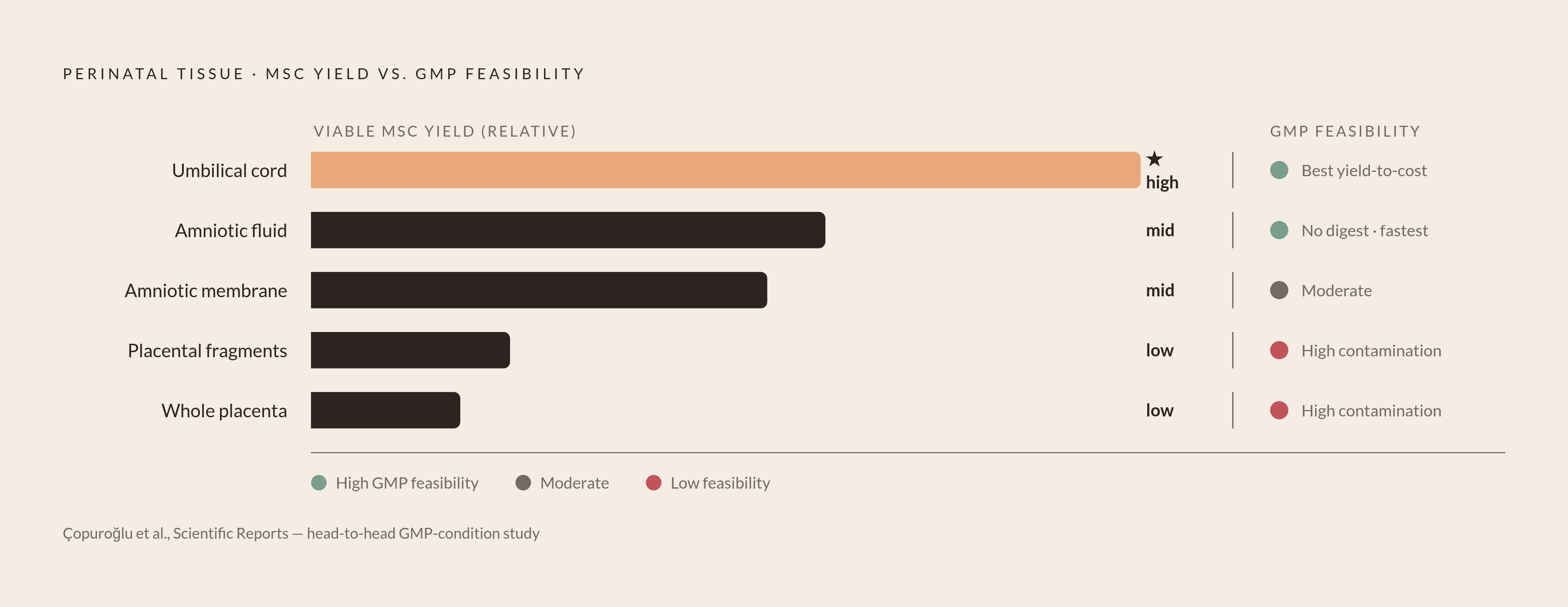
Under GMP conditions, the number that counts isn't total cells isolated but viable, sterile, identity-confirmed cells per unit of cost, and placental tissue as a whole does worse by that measure than several of the perinatal sources sitting right next to it, despite its biological richness.
Contamination drives much of that gap. Placental fragments and whole placenta show the highest contamination rates of any perinatal source under GMP conditions, which lengthens processing time and raises sterility costs in ways that quietly erode the economics of every cell produced. Amniotic fluid, by contrast, scored highest for feasibility in the Çopuroğlu et al. study, needed no enzymatic digestion step, and had the shortest median isolation time, making it the most GMP-efficient source even though it doesn't produce the highest raw yield. Umbilical cord delivered the best yield-to-cost ratio once normalized per million cells, a result that undercuts the common assumption that whole placenta's larger volume automatically makes it the better value at GMP scale.
A Phase 1 trial published in Scientific Reports, run by Shokati and colleagues at Shariati Hospital in Tehran, illustrates the GMP logistics in practice by manufacturing GMP-grade placenta-derived MSCs for patients with secondary progressive multiple sclerosis. The protocol required aseptic double-bagging of the placenta inside the operative suite itself, followed by transport in a cool box to the hospital's Research Institute for Oncology, Hematology & Cell Therapy Facility. That detail shows what GMP-grade placental harvest demands in physical infrastructure, coordination between surgical and laboratory teams, and chain-of-custody discipline separate from the biology of the cells themselves.
Open manual processing carries both a safety risk and a high labor cost that don't scale to routine commercial production, which is pushing the field toward closed-system and automated approaches instead. The Huasan Biotech protocol, with its sterile blender bags and fixed enzyme concentrations, represents one version of that shift: moving the main source of contamination risk away from individual operator technique and into the design of the system itself. That shift, more than any single yield figure, is what separates a promising placental cell source in a research paper from one that a manufacturer can actually produce, batch after batch, to a standard a regulator will accept.