Wharton's Jelly MSC Isolation Protocols and Yield Optimization
Precise dissection and enzyme choice determine stem cell yield from umbilical cord tissue.

Wharton's jelly, the gelatinous tissue packed around the umbilical vessels, has become one of the most practical sources of mesenchymal stem cells in regenerative medicine. The reason is simple: the cord gets thrown away after every birth, there's no invasive harvest and no ethical fight attached to it, and the tissue gives up cells in a way other sources just don't.
Bone marrow, the traditional MSC source, needs a needle into the hip and yields MSCs at a frequency of 0.001 to 0.01 percent of nucleated cells, a number that gets worse as the donor ages. Wharton's jelly doesn't have that problem. It takes up the largest cross-sectional area of the cord, and fresh isolation, no culture expansion needed, can pull roughly 4.61 ± 0.57 × 10⁶ cells per square centimeter. Amnion, subamnion, and perivascular fractions don't come close. Add in the low immunogenicity of these cells and their ability to turn into several different tissue types, and it makes sense that WJ-MSCs have become a manufacturing target and not just something people study in a lab for curiosity's sake.
None of that potential shows up on its own. It shows up only if every step of the isolation protocol, starting with how the tissue gets cut, gets handled right.
How the anatomy of the cord shapes what dissection can recover
Wharton's jelly isn't one uniform blob of matrix. It's connective tissue with sub-regions that differ in cell density and in how well those cells grow once you pull them out. Cells sitting close to the amniotic surface, inside the WJ layer, divide more readily than cells elsewhere in the matrix. So the very first cut a technician makes, whether it keeps that lining intact or wrecks it, has a direct bearing on how many usable cells come out the other end.
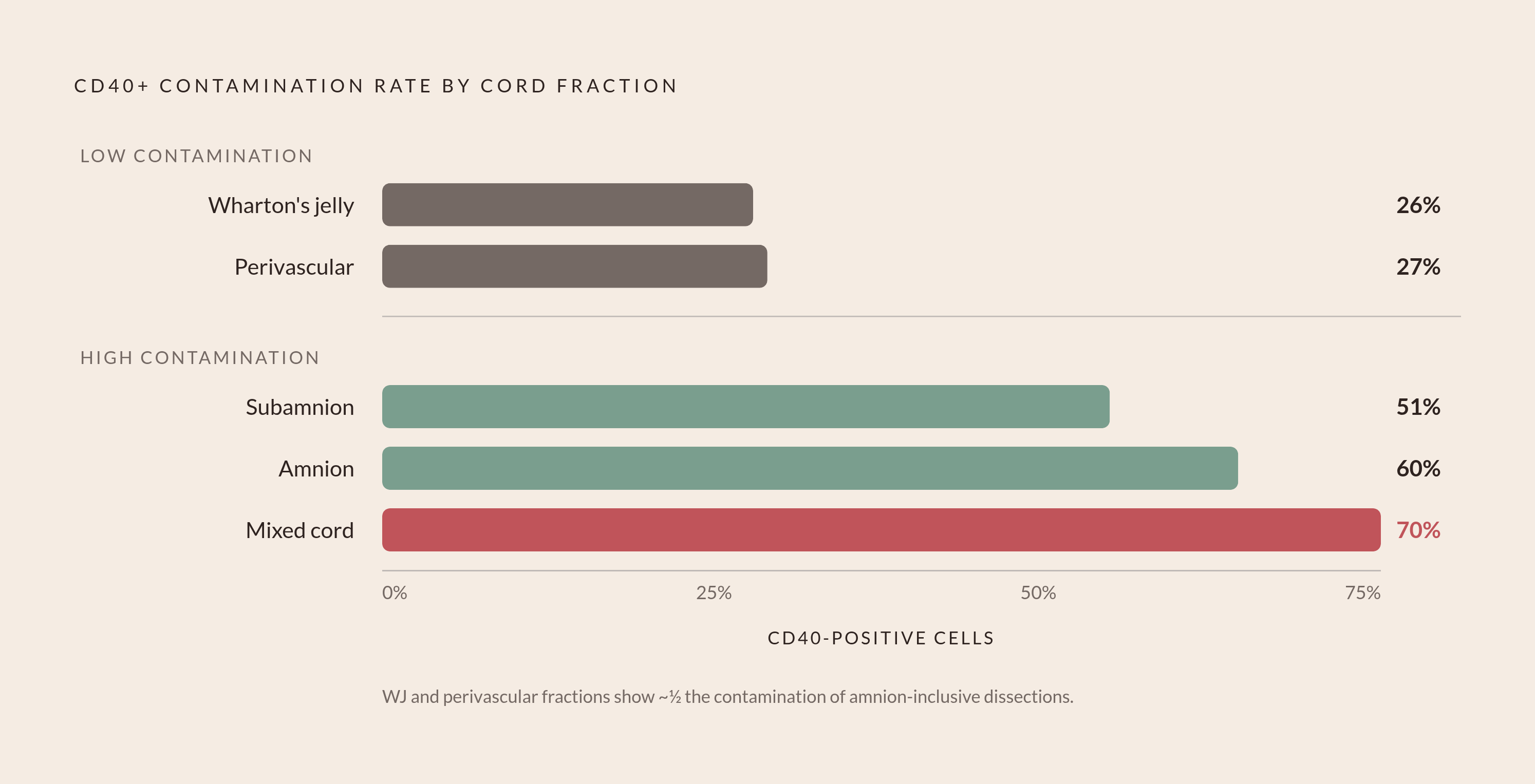
Purity follows the same pattern. CD40-positive cells, a marker for non-stem contaminating populations, show up at much lower rates in WJ and perivascular fractions (26 to 27 percent) than in subamnion, amnion, or mixed-cord fractions, where contamination runs from 51 to 70 percent. Where you dissect isn't just a yield question. It's a purity question from the first cut.
A 2024 technique published in JoVE takes this seriously. Instead of cutting the epithelial layer away with scissors, the protocol peels it lengthwise using mosquito clamps, a blunt dissection move meant to keep the subamniotic WJ layer in one piece. Sharp dissection risks tearing through or tossing out exactly the tissue where the high-proliferation cells live, and the reported result was better yield and better viability than conventional cutting produced.
Location within the cord matters just as much as depth. Explants taken from the cord-placenta junction start producing outgrowth in 3 to 4 days, segments from the length of the cord take 7 to 10 days, and explants from the fetal plate take longest of all, 11 to 14 days. Dissection isn't housekeeping before the real work starts. It decides how much cell material a lab has to work with and how long the wait for it runs.
Explant versus enzymatic digestion: what each method actually trades away
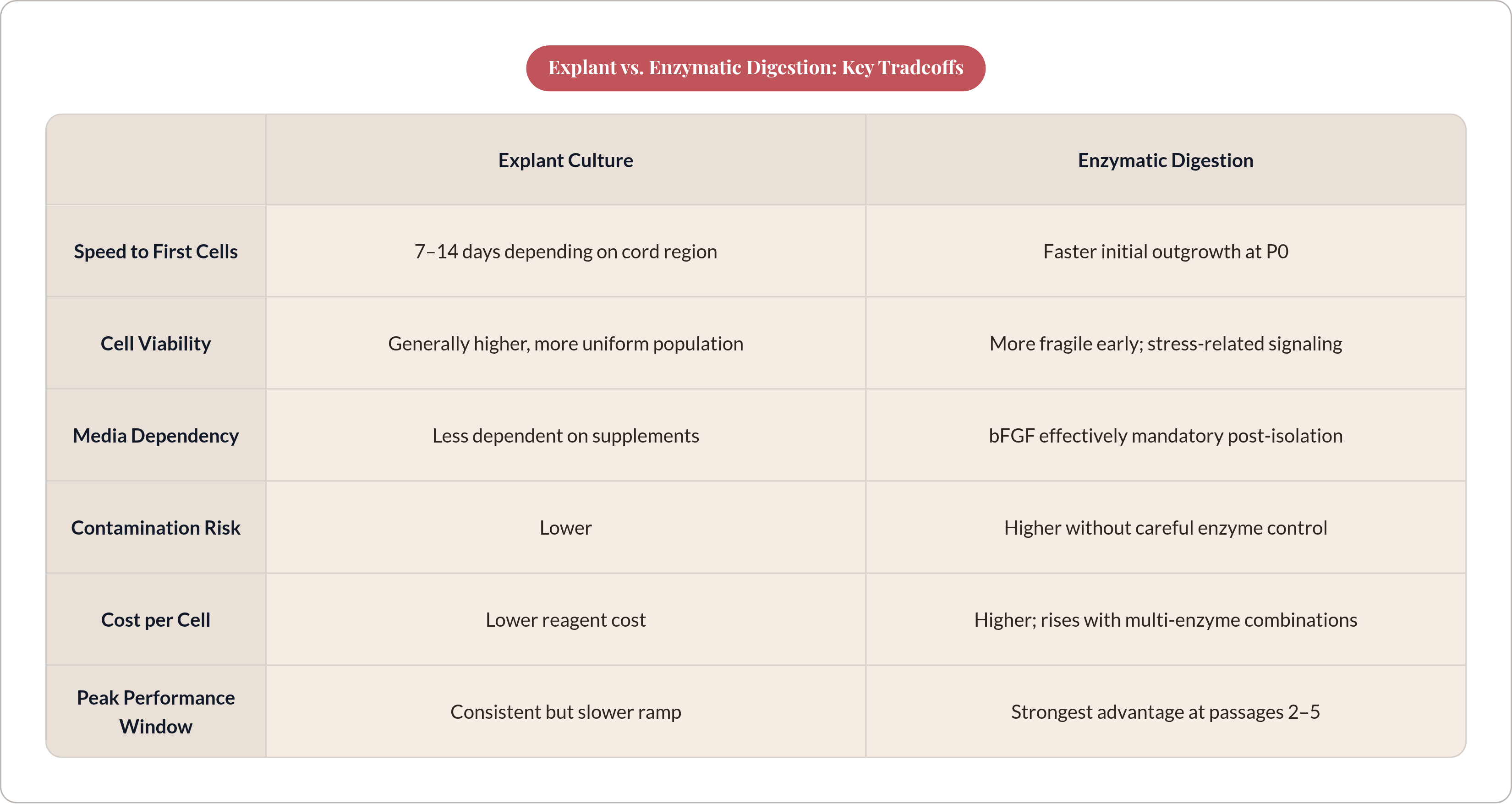
Four methods dominate the literature: a three-enzyme combination of collagenase, hyaluronidase, and trypsin (CHT), a two-enzyme collagenase-trypsin combination (CT), trypsin alone, and explant culture, where tissue fragments just sit on a surface and let cells migrate out on their own. All four produce WJ-MSCs, but not the same way, and not at the same cost. All four produce WJ-MSCs, but not the same way, and not at the same cost.
Explant culture skips proteolytic stress entirely. Cells keep their natural contact with the surrounding matrix as they migrate out, which helps them stick once they hit the culture surface, and the method carries lower contamination risk and a lower reagent bill than the enzymatic routes. The literature generally reports a more uniform, higher-viability population coming out of explant culture. Speed is the tradeoff: outgrowth from cord-length segments takes 7 to 10 days, and the whole isolation timeline stretches to match. In a six-method comparison published in Cell Transplantation (SAGE, 2022), two explant methods starting from 2 grams of WJ tissue each produced roughly 5 × 10⁵ cells after 14 days.
Enzymatic digestion trades patience for speed. Collagenase, sometimes paired with other enzymes, breaks the matrix down and dumps cells directly into suspension. In that same 2022 study, a two-step enzymatic protocol labeled M6 beat every other method on cell shape, yield, growth rate, and purity. That performance costs something, though: harsher multi-enzyme combinations chew through the extracellular meshwork that helps cells stick and survive, and the more enzyme-sensitive cells in the population get lost or damaged along the way. Multi-enzyme protocols also run more expensive per cell recovered than explant methods.
A PLOS ONE proteomic study found something that should worry anyone leaning on enzymatic digestion by default: without bFGF supplementation, the method caused early culture failure in two out of three donors tested. That single result reframes the whole comparison. Enzymatic digestion isn't just faster and harsher; it depends on downstream media support in a way explant culture doesn't. Neither method wins outright, and the choice made here sets constraints, particularly on media composition, that carry through every later stage of the protocol.
Optimizing enzymatic digestion: concentration, timing, and what the GMP literature has resolved
If a lab goes with enzymatic digestion, collagenase concentration becomes the lever that determines everything downstream. Too little enzyme and digestion stays incomplete; too much, and the cells take damage they may never recover from. A 2024 study in Stem Cell Research & Therapy worked through this tradeoff for GMP-grade production and landed on a specific answer: 0.4 PZ U/mL of Collagenase NB6, digested for 3 hours, produced the highest P0 yield of WJ-MSCs in their system.
Starting material matters here too, and not in a trivial way. The same study found a straight-line positive correlation between umbilical cord tissue weight and the number of P0 cells recovered. Tissue weight isn't a fixed given; it's a yield variable in its own right, one that varies with the donor before a lab technician ever picks up a scalpel.
The 2024 results also confirmed something the earlier method comparisons had already suggested: enzymatic digestion produces faster initial outgrowth at P0 than explant culture, and the viability and growth edge holds through passages 2 to 5. What single-enzyme optimization doesn't resolve is the tradeoff that comes with multi-enzyme combinations. CHT, CT, and collagenase alone each carry different levels of cell stress, and the 2022 six-method study's headline finding, that M6 outperformed the field, came with a caveat attached: cost and cell-sensitivity concerns are still live issues for anyone trying to run this at clinical scale.
For GMP teams, the 0.4 PZ U/mL, 3-hour parameter set is a floor, not a finished answer. Getting digestion right gets a lab a good starting cell number, though whether that number turns into a usable, scalable product still depends on what happens in culture after that.
How culture media composition (especially bFGF) determines whether isolated cells thrive or fail
Base media choice, α-MEM versus DMEM-F12, matters less than what gets added to either one. A 2024 study in the Avicenna Journal found that adding basic fibroblast growth factor (bFGF) to either base media significantly increased MSC proliferation compared to controls, while neither non-essential amino acids nor L-glutamine made a measurable difference on their own.
The PLOS ONE proteomic study builds directly on that. The highest viability observed across their comparison came from a non-scraped explant method supplemented with bFGF, and bFGF cut down cell doubling time regardless of isolation method. Proteomic profiling across all the groups tested found 2,372 proteins commonly expressed no matter how the cells were isolated or whether bFGF was present, a useful confirmation that core MSC identity doesn't shift with protocol. The pathway-level picture is more interesting: bFGF-treated cultures showed enrichment in oxidative phosphorylation and fatty acid metabolism pathways, while bFGF-free cultures and enzymatic-only groups showed more inflammatory and stress-related signaling. bFGF isn't just pushing cells to divide faster. It looks like it's actively damping the stress response that enzymatic digestion sets off in the first place.
Two of three donors failing in culture without bFGF after enzymatic isolation isn't a footnote; it's the finding that should change how a lab writes its media protocol. Once a lab commits to enzymatic digestion, bFGF stops being optional. It becomes close to mandatory, with real cost and formulation consequences attached.
Serum source matters just as much. Comparing human platelet lysate to fetal bovine serum in the same generation of culture, mean population doubling time ran roughly 23.5 hours with 5% hPL versus roughly 38.2 hours with 10% FBS, a real proliferative gap in hPL's favor. Per findings published at link.springer.com, 2% and 5% hPL concentrations produced similar expansion, so the cheaper, lower concentration gets the job done without giving up yield.
Passage timing and the window during which yield and quality are simultaneously high
Passage number gets treated too often as a simple harvest-time call, a matter of when the flask looks full enough. Passage timing interacts with isolation method, media formulation, and the quality of the starting cell population, so the "right" passage to harvest at isn't fixed. It's conditional on everything that happened before it.
A GMP optimization study tracking cells out to passage 9 found the viability and growth edge of enzymatic isolation over explant culture was strongest specifically across passages 2 through 5. Early on, at P0 and P1, enzymatic-derived cells grow out faster but stay more fragile; bFGF supplementation is what stabilizes them enough to keep expanding. Later passages bring their own risk, replicative senescence and drift away from the original phenotype, which is why the passage 2 to 5 window isn't just a convenient bucket. It's the performance peak the GMP-grade studies actually measured.
Whatever passage a team lands on for downstream use, characterization has to hold up at that exact point. The ISCT markers, CD73, CD90, CD105, and CD44, need to stay high (representative figures run 93.72%, 93.72%, 92.53%, and 98.55% respectively), and the hematopoietic markers CD34, CD14, and HLA-DR need to stay close to zero (3.96%, 3.15%, 0.57%). Scaling into a 3D bioreactor adds a wrinkle: CD105 can read as artificially low in 3D culture, simply because of the trypsin concentration used to strip cells off microcarriers. Teams need to confirm multipotency separately in that case, or they'll mistake a harvesting artifact for a real loss of stem cell character.
Any validated scale-up path, from lab flask to pilot-scale cell factory, only works if the passage timing decisions made at bench scale carry over intact. Cell factory volumes don't shrink the consequences of passaging too early or too late; they magnify them.
Variables that fall outside the standard protocol but materially affect yield
Cord tissue weight, already flagged as a driver of P0 yield, is itself downstream of things no protocol controls: donor variation, gestational age, maternal health factors. Some of the yield outcome gets locked in before a lab technician ever touches the tissue.
Cryopreservation is where the field has a real, unresolved disagreement, and it's worth sitting with rather than smoothing over. One study found that enzymatic digestion of cryopreserved WJ tissue produced the most viable MSCs of any method tested, beating both explant and mechanical digestion. Other reported protocols have found the opposite: isolation from frozen tissue fragments simply didn't work. Those two results contradict each other directly. The likeliest explanation is differences in cryoprotectant choice and tissue handling before freezing, not a flaw in either study, but that's an inference, not a settled fact. Teams running cord blood banking alongside WJ-MSC isolation shouldn't assume a protocol validated on fresh tissue will transfer to frozen tissue; it needs its own validation, run separately.
Compartment choice adds another layer. Subamnion, perivascular, and mixed-cord fractions all need a culture step before a lab can recover fresh cells, unlike WJ proper, which yields fresh cells directly. Which compartments a processing protocol includes changes both the total yield and the contamination profile of what comes out the other end.
Donor factors, gestational age, delivery mode, how long the cord sits before processing starts, are documented influences on yield in their own right. A protocol tuned and validated on a narrow donor population may simply not reproduce once it's run against a broader biobank population. Getting yield optimization right means pairing a well-characterized protocol with a defined donor-tissue specification, since the protocol variables and the biological variables don't sit apart from each other. They compound.
Integrating protocol decisions into a reproducible isolation workflow
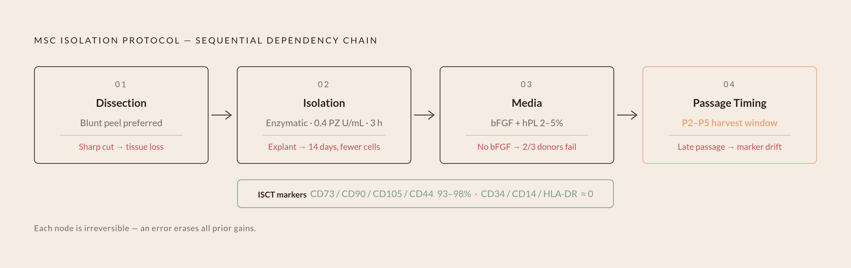
Dissection method determines which cells enter the process and in what shape. Isolation method, explant or enzymatic, and if enzymatic, at what concentration and for how long, determines the initial cell count and how much stress those cells carry into culture. Media composition, bFGF above all, plus serum source, determines whether the cells that survive isolation actually settle down and expand. Passage timing determines the point at which cells get pulled for use or for scale-up.
No single decision in that chain carries the whole result on its own. Enzymatic digestion run at the optimal 0.4 PZ U/mL, 3-hour parameter still underperforms if bFGF isn't in the media. Blunt dissection's advantage disappears if the enzymatic step that follows it runs too hot, and hPL media's proliferative edge gets erased if cells get harvested past the passage 5 window. Each decision only pays off if the ones around it were made correctly too.
Characterization isn't a final gate that cells pass or fail. It's the feedback loop that tells a team whether the upstream choices actually worked. The ISCT criteria, plastic adherence, positive expression of CD73, CD90, and CD105, negative expression of CD34 and CD45, and confirmed trilineage differentiation, function as the objective anchor for quality, not a box to check at the end.
For GMP and clinical manufacturing, the best-supported approach in the current literature pairs xenofree, hPL-based media with validated collagenase parameters and a harvest at passage 5 or earlier. Teams moving into cell factory-scale production should lock in passage decisions at bench scale first, before trying to amplify them. For biobanking operations working from cryopreserved starting material, the cryoprotectant protocol needs its own independent validation, not an assumption that whatever worked on fresh tissue will carry over.
The umbilical cord is already routinely available, already flowing through infrastructure that cord blood banks have spent years building and running. WJ-MSC isolation is a natural extension of that infrastructure, nothing more exotic than that. It only pays off, though, when every decision along the way, from the first cut to the last passage, gets made with the rest of the chain in mind.